 Movie
Movie Controller


+ Open data

- Basic information
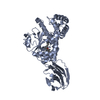
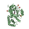
| Entry | Database: PDB / ID: 4omk | ||||||
|---|---|---|---|---|---|---|---|
| Title | Crystal structure of SPF bound to squalene | ||||||
 Components Components | SEC14-like protein 2 | ||||||
 Keywords Keywords |  TRANSPORT PROTEIN / cholesterol synthesis / squalene / 2 / 3-oxidosqualene / SEC14-like / CRAL-TRIO domain / hydrophobic ligand transporter TRANSPORT PROTEIN / cholesterol synthesis / squalene / 2 / 3-oxidosqualene / SEC14-like / CRAL-TRIO domain / hydrophobic ligand transporter | ||||||
| Function / homology |  Function and homology informationvitamin E binding / regulation of cholesterol biosynthetic process / phospholipid binding / positive regulation of DNA-templated transcription / extracellular exosome / nucleoplasm / nucleus / cytosol / cytoplasm Function and homology informationvitamin E binding / regulation of cholesterol biosynthetic process / phospholipid binding / positive regulation of DNA-templated transcription / extracellular exosome / nucleoplasm / nucleus / cytosol / cytoplasmSimilarity search - Function | ||||||
| Biological species |  Homo sapiens (human) Homo sapiens (human) | ||||||
| Method | X-RAY DIFFRACTION / SYNCHROTRON / MOLECULAR REPLACEMENT / Resolution: 1.75 Å | ||||||
 Authors Authors | Christen, M. / Marcaida, M.J. / Lamprakis, C. / Cascella, M. / Stocker, A. | ||||||
 Citation Citation | Journal: J.Struct.Biol. / Year: 2015 Title: Structural insights on cholesterol endosynthesis: Binding of squalene and 2,3-oxidosqualene to supernatant protein factor. Authors: Christen, M. / Marcaida, M.J. / Lamprakis, C. / Aeschimann, W. / Vaithilingam, J. / Schneider, P. / Hilbert, M. / Schneider, G. / Cascella, M. / Stocker, A. | ||||||
| History |
|
- Structure visualization
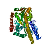
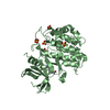
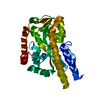
| Structure viewer | Molecule: MolmilJmol/JSmol |
|---|
- Downloads & links
-Download
| PDBx/mmCIF format | 4omk.cif.gz | 247.5 KB | Display | PDBx/mmCIF format |
|---|---|---|---|---|
| PDB format | pdb4omk.ent.gz | 201.8 KB | Display | PDB format |
| PDBx/mmJSON format | 4omk.json.gz | Tree view | PDBx/mmJSON format | |
| Others |  Other downloads Other downloads |
-Validation report
| Arichive directory | https://data.pdbj.org/pub/pdb/validation_reports/om/4omkftp://data.pdbj.org/pub/pdb/validation_reports/om/4omk | HTTPS FTP |
|---|
-Related structure data
| Related structure data |  4omjC  1o6uS S: Starting model for refinement C: citing same article ( |
|---|---|
| Similar structure data |


-Links
 PDBj
PDBj
- Assembly
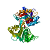
| Deposited unit | 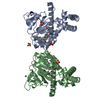
| ||||||||
|---|---|---|---|---|---|---|---|---|---|
| 1 | 
| ||||||||
| 2 | 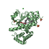
| ||||||||
| Unit cell |
|
-Components
-Protein , 1 types, 2 molecules AB
| #1: Protein | Mass: 31989.086 Da / Num. of mol.: 2 / Fragment: UNP residues 1-275 Source method: isolated from a genetically manipulated source Source: (gene. exp.) Homo sapiens (human) / Gene: C22orf6, KIAA1186, KIAA1658, SEC14L2 / Plasmid: pET28a / Production host:  Escherichia coli (E. coli) / Strain (production host): Lemo21 (DE3) / References: UniProt: O76054 Escherichia coli (E. coli) / Strain (production host): Lemo21 (DE3) / References: UniProt: O76054 |
|---|
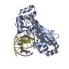
-Non-polymers , 5 types, 865 molecules 

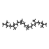


| #2: Chemical | ChemComp-SO4 / Sulfate Mass: 96.063 Da / Num. of mol.: 6 / Source method: obtained synthetically / Formula: SO4 Mass: 96.063 Da / Num. of mol.: 6 / Source method: obtained synthetically / Formula: SO4#3: Chemical | Chloride Mass: 35.453 Da / Num. of mol.: 2 / Source method: obtained synthetically / Formula: Cl Mass: 35.453 Da / Num. of mol.: 2 / Source method: obtained synthetically / Formula: Cl#4: Chemical | Squalene Mass: 410.718 Da / Num. of mol.: 2 / Source method: obtained synthetically / Formula: C30H50 Mass: 410.718 Da / Num. of mol.: 2 / Source method: obtained synthetically / Formula: C30H50#5: Chemical | Isopropyl alcohol Mass: 60.095 Da / Num. of mol.: 2 / Source method: obtained synthetically / Formula: C3H8O / Comment: alkaloid*YM Mass: 60.095 Da / Num. of mol.: 2 / Source method: obtained synthetically / Formula: C3H8O / Comment: alkaloid*YM#6: Water | ChemComp-HOH / | WaterMass: 18.015 Da / Num. of mol.: 853 / Source method: isolated from a natural source / Formula: H2O |
|---|
-Experimental details
-Experiment
| Experiment | Method: X-RAY DIFFRACTION / Number of used crystals: 1 |
|---|
- Sample preparation
| Crystal | Density Matthews: 3.83 Å3/Da / Density % sol: 67.9 % |
|---|---|
| Crystal grow | Temperature: 292 K / Method: vapor diffusion, hanging drop / pH: 6.5 Details: 0.2 M Lithium sulfate, 0.1 M Bis-Tris, 25% PEG3350, pH 6.5, VAPOR DIFFUSION, HANGING DROP, temperature 292K |
-Data collection
| Diffraction | Mean temperature: 100 K |
|---|---|
| Diffraction source | Source: SYNCHROTRON / Site: SLS  / Beamline: X06DA / Wavelength: 0.9999 Å / Beamline: X06DA / Wavelength: 0.9999 Å |
| Detector | Type: PSI PILATUS 6M / Detector: PIXEL / Date: Jun 21, 2012 |
| Radiation | Monochromator: Si (111) / Protocol: SINGLE WAVELENGTH / Monochromatic (M) / Laue (L): M / Scattering type: x-ray |
| Radiation wavelength | Wavelength: 0.9999 Å / Relative weight: 1 |
| Reflection | Resolution: 1.75→30 Å / Num. all: 97554 / Num. obs: 97261 / % possible obs: 99.7 % / Observed criterion σ(F): 2 / Observed criterion σ(I): 2 / Redundancy: 3.1 % / Rmerge(I) obs: 0.052 / Net I/σ(I): 12.7 |
| Reflection shell | Resolution: 1.75→1.84 Å / Redundancy: 3.3 % / Rmerge(I) obs: 0.317 / Mean I/σ(I) obs: 3.6 / Num. unique all: 14143 / % possible all: 99.8 |
- Processing
| Software |
| |||||||||||||||||||||||||||||||||||||||||||||||||||||||||||||||||||||||||||||||||||||||||||||||||||||||||||||||||||||||||||||||||||||||||||||||||||||||||||||||||||||||||||||||||||||||||||||||||||||||||||||||||||||||||
|---|---|---|---|---|---|---|---|---|---|---|---|---|---|---|---|---|---|---|---|---|---|---|---|---|---|---|---|---|---|---|---|---|---|---|---|---|---|---|---|---|---|---|---|---|---|---|---|---|---|---|---|---|---|---|---|---|---|---|---|---|---|---|---|---|---|---|---|---|---|---|---|---|---|---|---|---|---|---|---|---|---|---|---|---|---|---|---|---|---|---|---|---|---|---|---|---|---|---|---|---|---|---|---|---|---|---|---|---|---|---|---|---|---|---|---|---|---|---|---|---|---|---|---|---|---|---|---|---|---|---|---|---|---|---|---|---|---|---|---|---|---|---|---|---|---|---|---|---|---|---|---|---|---|---|---|---|---|---|---|---|---|---|---|---|---|---|---|---|---|---|---|---|---|---|---|---|---|---|---|---|---|---|---|---|---|---|---|---|---|---|---|---|---|---|---|---|---|---|---|---|---|---|---|---|---|---|---|---|---|---|---|---|---|---|---|---|---|---|
| Refinement | Method to determine structure: MOLECULAR REPLACEMENT Starting model: PDB entry 1O6U Resolution: 1.75→29.887 Å / SU ML: 0.17 / σ(F): 1.19 / Phase error: 17.26 / Stereochemistry target values: ML
| |||||||||||||||||||||||||||||||||||||||||||||||||||||||||||||||||||||||||||||||||||||||||||||||||||||||||||||||||||||||||||||||||||||||||||||||||||||||||||||||||||||||||||||||||||||||||||||||||||||||||||||||||||||||||
| Solvent computation | Shrinkage radii: 0.9 Å / VDW probe radii: 1.11 Å / Solvent model: FLAT BULK SOLVENT MODEL | |||||||||||||||||||||||||||||||||||||||||||||||||||||||||||||||||||||||||||||||||||||||||||||||||||||||||||||||||||||||||||||||||||||||||||||||||||||||||||||||||||||||||||||||||||||||||||||||||||||||||||||||||||||||||
| Refinement step | Cycle: LAST / Resolution: 1.75→29.887 Å
| |||||||||||||||||||||||||||||||||||||||||||||||||||||||||||||||||||||||||||||||||||||||||||||||||||||||||||||||||||||||||||||||||||||||||||||||||||||||||||||||||||||||||||||||||||||||||||||||||||||||||||||||||||||||||
| Refine LS restraints |
| |||||||||||||||||||||||||||||||||||||||||||||||||||||||||||||||||||||||||||||||||||||||||||||||||||||||||||||||||||||||||||||||||||||||||||||||||||||||||||||||||||||||||||||||||||||||||||||||||||||||||||||||||||||||||
| LS refinement shell |
|
