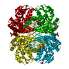
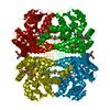
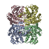
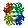
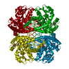
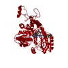
[English] 日本語
Yorodumi
- PDB-4oma: The crystal structure of methionine gamma-lyase from Citrobacter ... -

+
Open data


ID or keywords:

Loading...

-
Basic information

Entry
Database: PDB / ID: 4oma
TitleThe crystal structure of methionine gamma-lyase from Citrobacter freundii in complex with L-cycloserine pyridoxal-5'-phosphate
Componentsmethionine gamma-lyase
KeywordsLYASE / aminotransferase class I and II / methionine
Function / homology
Function and homology information


methionine gamma-lyase / methionine gamma-lyase activity / transsulfuration / pyridoxal phosphate binding
Similarity search - Function
L-methionine gamma-lyase / Cys/Met metabolism, pyridoxal phosphate-dependent enzyme / Cys/Met metabolism PLP-dependent enzyme / Aspartate Aminotransferase, domain 1 / Aspartate Aminotransferase, domain 1 / Aspartate Aminotransferase; domain 2 / Type I PLP-dependent aspartate aminotransferase-like (Major domain) / Pyridoxal phosphate-dependent transferase, small domain / Pyridoxal phosphate-dependent transferase, major domain / Pyridoxal phosphate-dependent transferase ...L-methionine gamma-lyase / Cys/Met metabolism, pyridoxal phosphate-dependent enzyme / Cys/Met metabolism PLP-dependent enzyme / Aspartate Aminotransferase, domain 1 / Aspartate Aminotransferase, domain 1 / Aspartate Aminotransferase; domain 2 / Type I PLP-dependent aspartate aminotransferase-like (Major domain) / Pyridoxal phosphate-dependent transferase, small domain / Pyridoxal phosphate-dependent transferase, major domain / Pyridoxal phosphate-dependent transferase / Alpha-Beta Complex / 3-Layer(aba) Sandwich / Alpha Beta
Similarity search - Domain/homology
Chem-LCS / DI(HYDROXYETHYL)ETHER / TRIETHYLENE GLYCOL / L-methionine gamma-lyase / :
Similarity search - Component
Biological speciesCitrobacter freundii (bacteria)
MethodX-RAY DIFFRACTION / SYNCHROTRON / MOLECULAR REPLACEMENT / Resolution: 1.6 Å
AuthorsRevtovich, S.V. / Nikulin, A.D. / Morozova, E.A. / Demidkina, T.V.
CitationJournal: J.Biol.Chem. / Year: 2015
Title: Pre-steady-state Kinetic and Structural Analysis of Interaction of Methionine gamma-Lyase from Citrobacter freundii with Inhibitors.
Authors: Kuznetsov, N.A. / Faleev, N.G. / Kuznetsova, A.A. / Morozova, E.A. / Revtovich, S.V. / Anufrieva, N.V. / Nikulin, A.D. / Fedorova, O.S. / Demidkina, T.V.
History
DepositionJan 27, 2014Deposition site: RCSB / Processing site: RCSB
Revision 1.0Nov 26, 2014Provider: repository / Type: Initial release
Revision 1.1Dec 3, 2014Group: Database references
Revision 1.2Jan 21, 2015Group: Database references
Revision 1.3Sep 20, 2023Group: Data collection / Database references ...Data collection / Database references / Derived calculations / Refinement description
Category: chem_comp_atom / chem_comp_bond ...chem_comp_atom / chem_comp_bond / database_2 / diffrn_source / pdbx_initial_refinement_model / struct_site
Item: _database_2.pdbx_DOI / _database_2.pdbx_database_accession ..._database_2.pdbx_DOI / _database_2.pdbx_database_accession / _diffrn_source.pdbx_synchrotron_site / _struct_site.pdbx_auth_asym_id / _struct_site.pdbx_auth_comp_id / _struct_site.pdbx_auth_seq_id

-
Structure visualization

Structure viewerMolecule:
MolmilJmol/JSmol

Downloads & links

-
Assembly

Deposited unit
A: methionine gamma-lyase
hetero molecules


Theoretical massNumber of molelcules
Total (without water)43,8167
Polymers42,9371
Non-polymers8796
Water4,792266
1
A: methionine gamma-lyase
hetero molecules

A: methionine gamma-lyase
hetero molecules

A: methionine gamma-lyase
hetero molecules

A: methionine gamma-lyase
hetero molecules


Theoretical massNumber of molelcules
Total (without water)175,26428
Polymers171,7474
Non-polymers3,51724
Water724
TypeNameSymmetry operationNumber
identity operation1_555x,y,z1
crystal symmetry operation2_555-x,-y,z1
crystal symmetry operation3_555-x,y,-z1
crystal symmetry operation4_555x,-y,-z1
Buried area27720 Å2
ΔGint-137 kcal/mol
Surface area46780 Å2
MethodPISA
Unit cell
Length a, b, c (Å)56.270, 122.890, 126.610
Angle α, β, γ (deg.)90.00, 90.00, 90.00
Int Tables number23
Space group name H-MI222
Components on special symmetry positions
IDModelComponents
11A-694-

HOH

21A-756-

HOH

31A-757-

HOH

-
Components

-
Protein , 1 types, 1 molecules A

#1: Protein methionine gamma-lyase /


Mass: 42936.801 Da / Num. of mol.: 1
Source method: isolated from a genetically manipulated source
Source: (gene. exp.) Citrobacter freundii (bacteria) / Gene: H922_06139, megl / Production host: Escherichia coli (E. coli) / Strain (production host): BL21(DE3)
References: UniProt: R1FPL7, UniProt: Q84AR1*PLUS, methionine gamma-lyase

-
Non-polymers , 5 types, 272 molecules

#2: Chemical ChemComp-LCS / [5-hydroxy-6-methyl-4-({[(4E)-3-oxo-1,2-oxazolidin-4-ylidene]amino}methyl)pyridin-3-yl]methyl dihydrogen phosphate


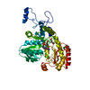
Mass: 331.219 Da / Num. of mol.: 1 / Source method: obtained synthetically / Formula: C11H14N3O7P
#3: Chemical ChemComp-PGE / TRIETHYLENE GLYCOL / Polyethylene glycol


Mass: 150.173 Da / Num. of mol.: 2 / Source method: obtained synthetically / Formula: C6H14O4
#4: Chemical ChemComp-PEG / DI(HYDROXYETHYL)ETHER / Diethylene glycol


Mass: 106.120 Da / Num. of mol.: 2 / Source method: obtained synthetically / Formula: C4H10O3
#5: Chemical ChemComp-CL / CHLORIDE ION / Chloride


Mass: 35.453 Da / Num. of mol.: 1 / Source method: obtained synthetically / Formula: Cl
#6: Water ChemComp-HOH / water / Water


Mass: 18.015 Da / Num. of mol.: 266 / Source method: isolated from a natural source / Formula: H2O

-
Experimental details

-
Experiment

ExperimentMethod: X-RAY DIFFRACTION / Number of used crystals: 1

-
Sample preparation

CrystalDensity Matthews: 2.55 Å3/Da / Density % sol: 51.74 %
Crystal growTemperature: 303 K / Method: vapor diffusion, hanging drop / pH: 8.5
Details: 5% PEG2000 MME, 50 mM Tris-HCl, 0.2 mM PLP, 0.25% DTT, pH 8.5, soaked in 50 mM L-cycloserine for 7 minutes, VAPOR DIFFUSION, HANGING DROP, temperature 303K

-
Data collection

DiffractionMean temperature: 100 K
Diffraction sourceSource: SYNCHROTRON / Site: EMBL/DESY, HAMBURG / Beamline: BW7B / Wavelength: 0.843 Å
DetectorType: MAR scanner 345 mm plate / Detector: IMAGE PLATE / Date: Nov 11, 2005
RadiationMonochromator: sagitally focused Si(111) / Protocol: SINGLE WAVELENGTH / Monochromatic (M) / Laue (L): M / Scattering type: x-ray
Radiation wavelengthWavelength: 0.843 Å / Relative weight: 1
ReflectionResolution: 1.6→15 Å / Num. all: 55080 / Num. obs: 55080 / % possible obs: 94.5 % / Observed criterion σ(F): 0 / Observed criterion σ(I): 0 / Redundancy: 4.5 % / Biso Wilson estimate: 26.8 Å2 / Rmerge(I) obs: 0.048 / Net I/σ(I): 17.28
Reflection shellResolution: 1.6→1.65 Å / Redundancy: 3.9 % / Rmerge(I) obs: 0.396 / Mean I/σ(I) obs: 3.6 / Num. unique all: 4606 / % possible all: 94.3

-
Processing

Software
NameVersionClassification
MAR345dtbdata collection
PHASESphasing
REFMAC5.8.0049refinement
PROCESSdata reduction
PROCESSdata scaling
RefinementMethod to determine structure: MOLECULAR REPLACEMENT
Starting model: PDB ENTRY 2RFV
Resolution: 1.6→15 Å / Cor.coef. Fo:Fc: 0.983 / Cor.coef. Fo:Fc free: 0.964 / SU B: 7.606 / SU ML: 0.102 / Cross valid method: THROUGHOUT / σ(F): 0 / σ(I): 0 / ESU R: 0.086 / ESU R Free: 0.081 / Stereochemistry target values: MAXIMUM LIKELIHOOD
RfactorNum. reflection% reflectionSelection details
Rfree0.197 2893 5.1 %RANDOM
Rwork0.139 ---
all0.142 53896 --
obs0.142 53896 97.48 %-
Solvent computationIon probe radii: 0.8 Å / Shrinkage radii: 0.8 Å / VDW probe radii: 1.2 Å / Solvent model: MASK
Displacement parametersBiso mean: 28.688 Å2
Baniso -1Baniso -2Baniso -3
1-5.51 Å2-0 Å2-0 Å2
2---2.81 Å20 Å2
3----2.7 Å2
Refine analyzeLuzzati coordinate error obs: 0.206 Å
Refinement stepCycle: LAST / Resolution: 1.6→15 Å
ProteinNucleic acidLigandSolventTotal
Num. atoms2993 0 57 266 3316
Refine LS restraints
Refine-IDTypeDev idealDev ideal targetNumber
X-RAY DIFFRACTIONr_bond_refined_d0.020.0193177
X-RAY DIFFRACTIONr_bond_other_d0.0030.023066
X-RAY DIFFRACTIONr_angle_refined_deg1.9431.9854314
X-RAY DIFFRACTIONr_angle_other_deg1.0143.0047060
X-RAY DIFFRACTIONr_dihedral_angle_1_deg6.355414
X-RAY DIFFRACTIONr_dihedral_angle_2_deg37.69624.308130
X-RAY DIFFRACTIONr_dihedral_angle_3_deg12.97915532
X-RAY DIFFRACTIONr_dihedral_angle_4_deg18.4271518
X-RAY DIFFRACTIONr_chiral_restr0.1440.2495
X-RAY DIFFRACTIONr_gen_planes_refined0.0090.0213592
X-RAY DIFFRACTIONr_gen_planes_other0.0010.02683
X-RAY DIFFRACTIONr_nbd_refined
X-RAY DIFFRACTIONr_nbd_other
X-RAY DIFFRACTIONr_nbtor_refined
X-RAY DIFFRACTIONr_nbtor_other
X-RAY DIFFRACTIONr_xyhbond_nbd_refined
X-RAY DIFFRACTIONr_xyhbond_nbd_other
X-RAY DIFFRACTIONr_metal_ion_refined
X-RAY DIFFRACTIONr_metal_ion_other
X-RAY DIFFRACTIONr_symmetry_vdw_refined
X-RAY DIFFRACTIONr_symmetry_vdw_other
X-RAY DIFFRACTIONr_symmetry_hbond_refined
X-RAY DIFFRACTIONr_symmetry_hbond_other
X-RAY DIFFRACTIONr_symmetry_metal_ion_refined
X-RAY DIFFRACTIONr_symmetry_metal_ion_other
X-RAY DIFFRACTIONr_mcbond_it4.7572.5961609
X-RAY DIFFRACTIONr_mcbond_other4.7382.5921605
X-RAY DIFFRACTIONr_mcangle_it5.9933.9122014
X-RAY DIFFRACTIONr_mcangle_other5.963.912013
X-RAY DIFFRACTIONr_scbond_it6.3763.0231566
X-RAY DIFFRACTIONr_scbond_other6.3743.0251567
X-RAY DIFFRACTIONr_scangle_it
X-RAY DIFFRACTIONr_scangle_other7.3644.372290
X-RAY DIFFRACTIONr_long_range_B_refined7.55522.7073784
X-RAY DIFFRACTIONr_long_range_B_other7.24822.2043676
X-RAY DIFFRACTIONr_rigid_bond_restr7.2636240
X-RAY DIFFRACTIONr_sphericity_free48.856564
X-RAY DIFFRACTIONr_sphericity_bonded22.14956381
LS refinement shellResolution: 1.6→1.642 Å / Total num. of bins used: 20
RfactorNum. reflection% reflection
Rfree0.293 200 -
Rwork0.275 3808 -
obs-4008 94.28 %

+
About Yorodumi

-
News

-
Feb 9, 2022. New format data for meta-information of EMDB entries

New format data for meta-information of EMDB entries

  • Version 3 of the EMDB header file is now the official format.
  • The previous official version 1.9 will be removed from the archive.

Related info.:EMDB header

External links:wwPDB to switch to version 3 of the EMDB data model

-
Aug 12, 2020. Covid-19 info

Covid-19 info

URL: https://pdbj.org/emnavi/covid19.php

New page: Covid-19 featured information page in EM Navigator.

Related info.:Covid-19 info / Mar 5, 2020. Novel coronavirus structure data

+
Mar 5, 2020. Novel coronavirus structure data

Novel coronavirus structure data

Related info.:Yorodumi Speices / Aug 12, 2020. Covid-19 info

External links:COVID-19 featured content - PDBj / Molecule of the Month (242):Coronavirus Proteases

+
Jan 31, 2019. EMDB accession codes are about to change! (news from PDBe EMDB page)

EMDB accession codes are about to change! (news from PDBe EMDB page)

  • The allocation of 4 digits for EMDB accession codes will soon come to an end. Whilst these codes will remain in use, new EMDB accession codes will include an additional digit and will expand incrementally as the available range of codes is exhausted. The current 4-digit format prefixed with “EMD-” (i.e. EMD-XXXX) will advance to a 5-digit format (i.e. EMD-XXXXX), and so on. It is currently estimated that the 4-digit codes will be depleted around Spring 2019, at which point the 5-digit format will come into force.
  • The EM Navigator/Yorodumi systems omit the EMD- prefix.

Related info.:Q: What is EMD? / ID/Accession-code notation in Yorodumi/EM Navigator

External links:EMDB Accession Codes are Changing Soon! / Contact to PDBj

+
Jul 12, 2017. Major update of PDB

Major update of PDB

  • wwPDB released updated PDB data conforming to the new PDBx/mmCIF dictionary.
  • This is a major update changing the version number from 4 to 5, and with Remediation, in which all the entries are updated.
  • In this update, many items about electron microscopy experimental information are reorganized (e.g. em_software).
  • Now, EM Navigator and Yorodumi are based on the updated data.

External links:wwPDB Remediation / Enriched Model Files Conforming to OneDep Data Standards Now Available in the PDB FTP Archive

-
Yorodumi

Thousand views of thousand structures

  • Yorodumi is a browser for structure data from EMDB, PDB, SASBDB, etc.
  • This page is also the successor to EM Navigator detail page, and also detail information page/front-end page for Omokage search.
  • The word "yorodu" (or yorozu) is an old Japanese word meaning "ten thousand". "mi" (miru) is to see.

Related info.:EMDB / PDB / SASBDB / Comparison of 3 databanks / Yorodumi Search / Aug 31, 2016. New EM Navigator & Yorodumi / Yorodumi Papers / Jmol/JSmol / Function and homology information / Changes in new EM Navigator and Yorodumi

Read more