Mass: 18.015 Da / Num. of mol.: 177 / Source method: isolated from a natural source / Formula: H2O
Sequence details
THE RNA STRAND BETWEEN RESIDUES A9 AND U21 IS DISORDERED. BOTH THE SEQUENCE AND LENGTH OF THE ...THE RNA STRAND BETWEEN RESIDUES A9 AND U21 IS DISORDERED. BOTH THE SEQUENCE AND LENGTH OF THE DISORDERED REGION ARE UNKNOWN.
-
Experimental details
-
Experiment
Experiment
Method: X-RAY DIFFRACTION / Number of used crystals: 1
-
Sample preparation
Crystal
Density Matthews: 2.21 Å3/Da / Density % sol: 44.43 %
Crystal grow
Temperature: 293 K / Method: vapor diffusion, hanging drop / pH: 9 Details: 16% PEG3350, 12% isopropanol, 0.1 M phenol, 0.1 M Tris, pH 9.0, VAPOR DIFFUSION, HANGING DROP, temperature 293.0K
-
Data collection
Diffraction
ID
Mean temperature (K)
Crystal-ID
1
100
1
2
100
1
Diffraction source
Source
Site
Beamline
ID
Wavelength (Å)
SYNCHROTRON
SSRL
BL11-1
1
0.97926, 0.91837
SYNCHROTRON
APS
24-ID-E
2
0.9792
Detector
Type
ID
Detector
Date
DECTRIS PILATUS 6M
1
PIXEL
Dec 9, 2011
ADSC QUANTUM 315
2
CCD
Nov 19, 2011
Radiation
ID
Monochromator
Protocol
Monochromatic (M) / Laue (L)
Scattering type
Wavelength-ID
1
Si(111)
MAD
M
x-ray
1
2
Si(111)
SINGLEWAVELENGTH
M
x-ray
1
Radiation wavelength
ID
Wavelength (Å)
Relative weight
1
0.97926
1
2
0.91837
1
3
0.9792
1
Reflection
Resolution: 2.2→65.493 Å / Num. obs: 44375 / % possible obs: 99.4 % / Redundancy: 3.8 % / Rmerge(I) obs: 0.057 / Χ2: 0.76 / Net I/σ(I): 11.5
Reflection shell
Resolution (Å)
Redundancy (%)
Rmerge(I) obs
Num. unique all
Χ2
Diffraction-ID
% possible all
2.2-2.28
3.7
0.741
4372
0.843
1,2
98.7
2.28-2.37
3.8
0.551
4413
0.843
1,2
98.9
2.37-2.48
3.8
0.386
4394
0.89
1,2
99.3
2.48-2.61
3.8
0.268
4454
0.959
1,2
99.3
2.61-2.77
3.8
0.174
4420
0.865
1,2
99.3
2.77-2.99
3.8
0.105
4434
0.846
1,2
99.7
2.99-3.29
3.8
0.063
4453
0.77
1,2
99.6
3.29-3.76
3.8
0.047
4444
0.712
1,2
99.7
3.76-4.74
3.8
0.045
4469
0.665
1,2
99.8
4.74-65.493
3.7
0.025
4522
0.205
1,2
99.3
-
Processing
Software
Name
Version
Classification
ADSC
Quantum
datacollection
PHENIX
modelbuilding
PHENIX
(phenix.refine: 1.8.2_1309)
refinement
HKL-2000
datareduction
HKL-2000
datascaling
PHENIX
phasing
Refinement
Method to determine structure: MAD / Resolution: 2.3→41.588 Å / SU ML: 0.34 / σ(F): 1.36 / Phase error: 27.81 / Stereochemistry target values: ML
Rfactor
Num. reflection
% reflection
Rfree
0.2529
1960
5.06 %
Rwork
0.212
-
-
obs
0.2141
38697
99.36 %
Solvent computation
Shrinkage radii: 0.9 Å / VDW probe radii: 1.11 Å / Solvent model: FLAT BULK SOLVENT MODEL
Refinement step
Cycle: LAST / Resolution: 2.3→41.588 Å
Protein
Nucleic acid
Ligand
Solvent
Total
Num. atoms
6343
191
11
177
6722
Refine LS restraints
Refine-ID
Type
Dev ideal
Number
X-RAY DIFFRACTION
f_bond_d
0.003
6711
X-RAY DIFFRACTION
f_angle_d
0.776
9117
X-RAY DIFFRACTION
f_dihedral_angle_d
14.352
2552
X-RAY DIFFRACTION
f_chiral_restr
0.053
1022
X-RAY DIFFRACTION
f_plane_restr
0.003
1138
LS refinement shell
Resolution (Å)
Rfactor Rfree
Num. reflection Rfree
Rfactor Rwork
Num. reflection Rwork
Refine-ID
% reflection obs (%)
2.3-2.3575
0.3449
137
0.2584
2615
X-RAY DIFFRACTION
99
2.3575-2.4213
0.3597
139
0.2566
2597
X-RAY DIFFRACTION
99
2.4213-2.4925
0.3125
144
0.2429
2598
X-RAY DIFFRACTION
99
2.4925-2.5729
0.3424
137
0.2351
2592
X-RAY DIFFRACTION
99
2.5729-2.6649
0.2819
146
0.238
2624
X-RAY DIFFRACTION
99
2.6649-2.7716
0.2976
139
0.2349
2621
X-RAY DIFFRACTION
99
2.7716-2.8977
0.268
142
0.2332
2633
X-RAY DIFFRACTION
100
2.8977-3.0504
0.3112
132
0.2401
2612
X-RAY DIFFRACTION
100
3.0504-3.2415
0.2991
157
0.2324
2611
X-RAY DIFFRACTION
100
3.2415-3.4916
0.2868
125
0.2295
2658
X-RAY DIFFRACTION
100
3.4916-3.8428
0.2276
120
0.2049
2633
X-RAY DIFFRACTION
100
3.8428-4.3983
0.2301
151
0.1907
2648
X-RAY DIFFRACTION
100
4.3983-5.5393
0.2032
141
0.1813
2648
X-RAY DIFFRACTION
100
5.5393-41.5946
0.2086
150
0.1987
2647
X-RAY DIFFRACTION
98
+
About Yorodumi
-
News
-
Feb 9, 2022. New format data for meta-information of EMDB entries
New format data for meta-information of EMDB entries
Version 3 of the EMDB header file is now the official format.
The previous official version 1.9 will be removed from the archive.
In the structure databanks used in Yorodumi, some data are registered as the other names, "COVID-19 virus" and "2019-nCoV". Here are the details of the virus and the list of structure data.
Jan 31, 2019. EMDB accession codes are about to change! (news from PDBe EMDB page)
EMDB accession codes are about to change! (news from PDBe EMDB page)
The allocation of 4 digits for EMDB accession codes will soon come to an end. Whilst these codes will remain in use, new EMDB accession codes will include an additional digit and will expand incrementally as the available range of codes is exhausted. The current 4-digit format prefixed with “EMD-” (i.e. EMD-XXXX) will advance to a 5-digit format (i.e. EMD-XXXXX), and so on. It is currently estimated that the 4-digit codes will be depleted around Spring 2019, at which point the 5-digit format will come into force.
The EM Navigator/Yorodumi systems omit the EMD- prefix.
Related info.:Q: What is EMD? / ID/Accession-code notation in Yorodumi/EM Navigator
Yorodumi is a browser for structure data from EMDB, PDB, SASBDB, etc.
This page is also the successor to EM Navigator detail page, and also detail information page/front-end page for Omokage search.
The word "yorodu" (or yorozu) is an old Japanese word meaning "ten thousand". "mi" (miru) is to see.
Related info.:EMDB / PDB / SASBDB / Comparison of 3 databanks / Yorodumi Search / Aug 31, 2016. New EM Navigator & Yorodumi / Yorodumi Papers / Jmol/JSmol / Function and homology information / Changes in new EM Navigator and Yorodumi
 Movie
Movie Controller


 Open data
 Basic information
 Components
 Keywords
 RNA-binding protein /
  Function and homology information
 Authors
 Citation
 Structure visualization
 Downloads & links
 Other downloads


 PDBj
PDBj


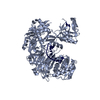
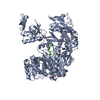
 Assembly
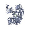


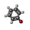
 Mass: 94.111 Da / Num. of mol.: 1 / Source method: obtained synthetically / Formula: C6H6O
 Mass: 60.095 Da / Num. of mol.: 1 / Source method: obtained synthetically / Formula: C3H8O / Comment: alkaloid*YM
 Mass: 18.015 Da / Num. of mol.: 177 / Source method: isolated from a natural source / Formula: H2O
 Sample preparation
 Processing