 Movie
Movie Controller


+ Open data

- Basic information
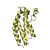
| Entry | Database: PDB / ID: 4ns0 | ||||||
|---|---|---|---|---|---|---|---|
| Title | The C2A domain of Rabphilin 3A in complex with PI(4,5)P2 | ||||||
 Components Components | Rabphilin-3A | ||||||
 Keywords Keywords |  PROTEIN TRANSPORT / C2 domain / Calcium binding / phospholipid binding / RABPHILIN-3A / C2A / SYNAPTIC EXOCYTOSIS METAL-BINDING / C-2 domain fold / EXOPHILIN-1 PROTEIN TRANSPORT / C2 domain / Calcium binding / phospholipid binding / RABPHILIN-3A / C2A / SYNAPTIC EXOCYTOSIS METAL-BINDING / C-2 domain fold / EXOPHILIN-1 | ||||||
| Function / homology |  Function and homology informationselenium binding / spontaneous neurotransmitter secretion / extrinsic component of synaptic vesicle membrane / regulation of calcium ion-dependent exocytosis / cholinergic synapse / inositol 1,4,5 trisphosphate binding / calcium-dependent phospholipid binding / dendritic spine organization / synaptic vesicle priming / extrinsic component of membrane ...selenium binding / spontaneous neurotransmitter secretion / extrinsic component of synaptic vesicle membrane / regulation of calcium ion-dependent exocytosis / cholinergic synapse / inositol 1,4,5 trisphosphate binding / calcium-dependent phospholipid binding / dendritic spine organization / synaptic vesicle priming / extrinsic component of membrane / phosphate ion binding / exocytosis / regulation of NMDA receptor activity / phosphatidylinositol-4,5-bisphosphate binding / secretory granule / phospholipid binding / intracellular protein transport / neuromuscular junction / synaptic vesicle membrane / small GTPase binding / synaptic vesicle / postsynaptic membrane / dendritic spine / neuron projection / synapse / calcium ion binding / protein-containing complex binding / protein-containing complex / zinc ion binding Function and homology informationselenium binding / spontaneous neurotransmitter secretion / extrinsic component of synaptic vesicle membrane / regulation of calcium ion-dependent exocytosis / cholinergic synapse / inositol 1,4,5 trisphosphate binding / calcium-dependent phospholipid binding / dendritic spine organization / synaptic vesicle priming / extrinsic component of membrane ...selenium binding / spontaneous neurotransmitter secretion / extrinsic component of synaptic vesicle membrane / regulation of calcium ion-dependent exocytosis / cholinergic synapse / inositol 1,4,5 trisphosphate binding / calcium-dependent phospholipid binding / dendritic spine organization / synaptic vesicle priming / extrinsic component of membrane / phosphate ion binding / exocytosis / regulation of NMDA receptor activity / phosphatidylinositol-4,5-bisphosphate binding / secretory granule / phospholipid binding / intracellular protein transport / neuromuscular junction / synaptic vesicle membrane / small GTPase binding / synaptic vesicle / postsynaptic membrane / dendritic spine / neuron projection / synapse / calcium ion binding / protein-containing complex binding / protein-containing complex / zinc ion bindingSimilarity search - Function | ||||||
| Biological species |  Rattus norvegicus (Norway rat) Rattus norvegicus (Norway rat) | ||||||
| Method | X-RAY DIFFRACTION / SYNCHROTRON / MOLECULAR REPLACEMENT / Resolution: 1.8 Å | ||||||
 Authors Authors | Guillen, J. / Ferrer-Orta, C. / Buxaderas, M. / Perez-sanchez, D. / Guerrero-Valero, M. / Luengo-Gil, G. / Pous, J. / Guerra, P. / Gomez-Fernandez, J.C. / Verdaguer, N. / Corbalan-Garcia, S. | ||||||
 Citation Citation | Journal: Proc.Natl.Acad.Sci.USA / Year: 2013 Title: Structural insights into the Ca2+ and PI(4,5)P2 binding modes of the C2 domains of rabphilin 3A and synaptotagmin 1. Authors: Guillen, J. / Ferrer-Orta, C. / Buxaderas, M. / Perez-Sanchez, D. / Guerrero-Valero, M. / Luengo-Gil, G. / Pous, J. / Guerra, P. / Gomez-Fernandez, J.C. / Verdaguer, N. / Corbalan-Garcia, S. | ||||||
| History |
|
- Structure visualization
| Structure viewer | Molecule: MolmilJmol/JSmol |
|---|
- Downloads & links
-Download
| PDBx/mmCIF format | 4ns0.cif.gz | 44.8 KB | Display | PDBx/mmCIF format |
|---|---|---|---|---|
| PDB format | pdb4ns0.ent.gz | 29.5 KB | Display | PDB format |
| PDBx/mmJSON format | 4ns0.json.gz | Tree view | PDBx/mmJSON format | |
| Others |  Other downloads Other downloads |
-Validation report
| Arichive directory | https://data.pdbj.org/pub/pdb/validation_reports/ns/4ns0ftp://data.pdbj.org/pub/pdb/validation_reports/ns/4ns0 | HTTPS FTP |
|---|
-Related structure data
| Related structure data |  4lt7C 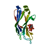 4np9C  2chdS C: citing same article ( S: Starting model for refinement |
|---|---|
| Similar structure data |


-Links
 PDBj
PDBj


- Assembly
| Deposited unit | 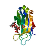
| ||||||||
|---|---|---|---|---|---|---|---|---|---|
| 1 |
| ||||||||
| Unit cell |
|
-Components
| #1: Protein | Mass: 15305.478 Da / Num. of mol.: 1 / Fragment: C2 domain, UNP RESIDUES 378-510 Source method: isolated from a genetically manipulated source Source: (gene. exp.) Rattus norvegicus (Norway rat) / Gene: Rph3a / Production host:  Escherichia coli (E. coli) / References: UniProt: P47709 Escherichia coli (E. coli) / References: UniProt: P47709 |
|---|---|
| #2: Chemical | ChemComp-PIO / [(  Mass: 746.566 Da / Num. of mol.: 1 / Source method: obtained synthetically / Formula: C25H49O19P3 Mass: 746.566 Da / Num. of mol.: 1 / Source method: obtained synthetically / Formula: C25H49O19P3 |
| #3: Chemical | ChemComp-SO4 / Sulfate  Mass: 96.063 Da / Num. of mol.: 1 / Source method: obtained synthetically / Formula: SO4 Mass: 96.063 Da / Num. of mol.: 1 / Source method: obtained synthetically / Formula: SO4 |
| #4: Water | ChemComp-HOH / Water Mass: 18.015 Da / Num. of mol.: 113 / Source method: isolated from a natural source / Formula: H2O Mass: 18.015 Da / Num. of mol.: 113 / Source method: isolated from a natural source / Formula: H2O |
-Experimental details
-Experiment
| Experiment | Method: X-RAY DIFFRACTION / Number of used crystals: 2 |
|---|
- Sample preparation
| Crystal | Density Matthews: 2.14 Å3/Da / Density % sol: 42.55 % |
|---|---|
| Crystal grow | Temperature: 293 K / Method: vapor diffusion, sitting drop / pH: 7.5 Details: 25mM Hepes, 20mM Ammonuim Sulphate, 25% PEG 4K, pH 7.5, VAPOR DIFFUSION, SITTING DROP, temperature 293K |
-Data collection
| Diffraction | Mean temperature: 100 K |
|---|---|
| Diffraction source | Source: SYNCHROTRON / Site: SOLEIL  / Beamline: PROXIMA 1 / Wavelength: 0.93 / Beamline: PROXIMA 1 / Wavelength: 0.93 |
| Detector | Type: PSI PILATUS 6M / Detector: PIXEL / Date: Mar 18, 2012 |
| Radiation | Protocol: SINGLE WAVELENGTH / Monochromatic (M) / Laue (L): M / Scattering type: x-ray |
| Radiation wavelength | Wavelength: 0.93 Å / Relative weight: 1 |
| Reflection | Resolution: 1.8→44.36 Å / Num. obs: 12421 / % possible obs: 94.2 % / Observed criterion σ(I): 1 / Redundancy: 2.9 % / Rmerge(I) obs: 0.131 / Rsym value: 0.061 / Net I/σ(I): 15 |
| Reflection shell | Resolution: 1.78→1.89 Å / Rmerge(I) obs: 0.72 / % possible all: 96.9 |
- Processing
| Software |
| ||||||||||||||||||||||||||||||||||||||||||||||||||||||||||||||||||||||||||||||||||||||||||||||||||||||||||||||||||||||||||||||||||||||||||||||||||||||||||||||||||||||||||||||||||||||
|---|---|---|---|---|---|---|---|---|---|---|---|---|---|---|---|---|---|---|---|---|---|---|---|---|---|---|---|---|---|---|---|---|---|---|---|---|---|---|---|---|---|---|---|---|---|---|---|---|---|---|---|---|---|---|---|---|---|---|---|---|---|---|---|---|---|---|---|---|---|---|---|---|---|---|---|---|---|---|---|---|---|---|---|---|---|---|---|---|---|---|---|---|---|---|---|---|---|---|---|---|---|---|---|---|---|---|---|---|---|---|---|---|---|---|---|---|---|---|---|---|---|---|---|---|---|---|---|---|---|---|---|---|---|---|---|---|---|---|---|---|---|---|---|---|---|---|---|---|---|---|---|---|---|---|---|---|---|---|---|---|---|---|---|---|---|---|---|---|---|---|---|---|---|---|---|---|---|---|---|---|---|---|---|
| Refinement | Method to determine structure: MOLECULAR REPLACEMENT Starting model: 2CHD Resolution: 1.8→44.36 Å / Cor.coef. Fo:Fc: 0.959 / Cor.coef. Fo:Fc free: 0.953 / SU B: 3.744 / SU ML: 0.1 / Cross valid method: THROUGHOUT / ESU R: 0.135 / ESU R Free: 0.129 / Stereochemistry target values: MAXIMUM LIKELIHOOD / Details: HYDROGENS HAVE BEEN ADDED IN THE RIDING POSITIONS
| ||||||||||||||||||||||||||||||||||||||||||||||||||||||||||||||||||||||||||||||||||||||||||||||||||||||||||||||||||||||||||||||||||||||||||||||||||||||||||||||||||||||||||||||||||||||
| Solvent computation | Ion probe radii: 0.8 Å / Shrinkage radii: 0.8 Å / VDW probe radii: 1.2 Å / Solvent model: MASK | ||||||||||||||||||||||||||||||||||||||||||||||||||||||||||||||||||||||||||||||||||||||||||||||||||||||||||||||||||||||||||||||||||||||||||||||||||||||||||||||||||||||||||||||||||||||
| Displacement parameters | Biso mean: 24.048 Å2
| ||||||||||||||||||||||||||||||||||||||||||||||||||||||||||||||||||||||||||||||||||||||||||||||||||||||||||||||||||||||||||||||||||||||||||||||||||||||||||||||||||||||||||||||||||||||
| Refinement step | Cycle: LAST / Resolution: 1.8→44.36 Å
| ||||||||||||||||||||||||||||||||||||||||||||||||||||||||||||||||||||||||||||||||||||||||||||||||||||||||||||||||||||||||||||||||||||||||||||||||||||||||||||||||||||||||||||||||||||||
| Refine LS restraints |
|