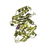
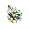
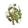
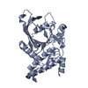
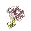
Entry Database : PDB / ID : 4nndTitle Structural basis of PTPN18 fingerprint on distinct HER2 tyrosine phosphorylation sites Receptor tyrosine-protein kinase erbB-2 Tyrosine-protein phosphatase non-receptor type 18 Keywords / / / / Function / homology Function Domain/homology Component
/ / / / / / / / / / / / / / / / / / / / / / / / / / / / / / / / / / / / / / / / / / / / / / / / / / / / / / / / / / / / / / / / / / / / / / / / / / / / / / / / / / / / / / / / / / / / / / / / / / / / / / / / / / / / / / / / / / / / / / / / / / / / / / / / / / / / / / / / / / / / / / / / / / / / / / / / / / / / / Biological species Homo sapiens (human)Method / / / Resolution : 2.502 Å Authors Wang, H. / Yang, F. / Yang, D. / Du, Y. #1: Journal : Cell(Cambridge,Mass.) / Year : 2009Title : Large-scale structural analysis of the classical human protein tyrosine phosphatome.
Authors :
Barr, A.J. / Ugochukwu, E. / Lee, W.H. / King, O.N. / Filippakopoulos, P. / Alfano, I. / Savitsky, P. / Burgess-Brown, N.A. / Muller, S. / Knapp, S. History Deposition Nov 17, 2013 Deposition site / Processing site Revision 1.0 Nov 19, 2014 Provider / Type
 Movie
Movie Controller

 Yorodumi
 Open data
 Basic information
 Components
 Keywords
 HYDROLASE /
  Function and homology information
 Authors
 Citation
 Structure visualization
 Downloads & links
 Other downloads
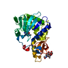


 PDBj
PDBj


 Assembly
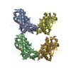


 Mass: 18.015 Da / Num. of mol.: 74 / Source method: isolated from a natural source / Formula: H2O
 Sample preparation
 / Beamline: BL17U / Wavelength: 1 Å
 Processing