 Movie
Movie Controller


+ Open data

- Basic information
| Entry | Database: PDB / ID: 4lwp | ||||||
|---|---|---|---|---|---|---|---|
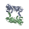
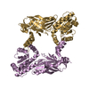
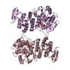
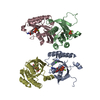
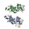
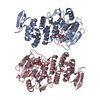
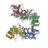
| Title | Crystal structure of PRMT6-SAH | ||||||
 Components Components | Arginine N-methyltransferase, putative | ||||||
 Keywords Keywords |  TRANSFERASE / SAM binding domain / arginine methylation TRANSFERASE / SAM binding domain / arginine methylation | ||||||
| Function / homology |  Function and homology information Function and homology informationprotein-arginine C-methyltransferase activity / : / peptidyl-arginine methylation, to asymmetrical-dimethyl arginine / histone arginine N-methyltransferase activity / cytosolSimilarity search - Function | ||||||
| Biological species |  Trypanosoma brucei brucei (eukaryote) Trypanosoma brucei brucei (eukaryote) | ||||||
| Method | X-RAY DIFFRACTION / SAD / Resolution: 2.353 Å | ||||||
 Authors Authors | Zhu, Y. / Wang, C. / Shi, Y. / Teng, M. | ||||||
 Citation Citation | Journal: Plos One / Year: 2014 Title: Crystal Structure of Arginine Methyltransferase 6 from Trypanosoma brucei Authors: Wang, C. / Zhu, Y. / Chen, J. / Li, X. / Peng, J. / Chen, J. / Zou, Y. / Zhang, Z. / Jin, H. / Yang, P. / Wu, J. / Niu, L. / Gong, Q. / Teng, M. / Shi, Y. | ||||||
| History |
|
- Structure visualization
| Structure viewer | Molecule: MolmilJmol/JSmol |
|---|
- Downloads & links
-Download
| PDBx/mmCIF format | 4lwp.cif.gz | 142.8 KB | Display | PDBx/mmCIF format |
|---|---|---|---|---|
| PDB format | pdb4lwp.ent.gz | 111 KB | Display | PDB format |
| PDBx/mmJSON format | 4lwp.json.gz | Tree view | PDBx/mmJSON format | |
| Others |  Other downloads Other downloads |
-Validation report
| Arichive directory | https://data.pdbj.org/pub/pdb/validation_reports/lw/4lwpftp://data.pdbj.org/pub/pdb/validation_reports/lw/4lwp | HTTPS FTP |
|---|
-Related structure data


-Links
 PDBj
PDBj- Assembly
| Deposited unit | 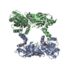
| |||||||||
|---|---|---|---|---|---|---|---|---|---|---|
| 1 |
| |||||||||
| Unit cell |
| |||||||||
| Components on special symmetry positions |
|
-Components
| #1: Protein | Mass: 41257.465 Da / Num. of mol.: 2 Source method: isolated from a genetically manipulated source Source: (gene. exp.) Trypanosoma brucei brucei (eukaryote) / Strain: 927/4 GUTat10.1 / Gene: Tb927.5.3960 / Production host:  Escherichia coli (E. coli) / References: UniProt: Q57U70 Escherichia coli (E. coli) / References: UniProt: Q57U70#2: Chemical | ChemComp-IOD / Iodide  Mass: 126.904 Da / Num. of mol.: 9 / Source method: obtained synthetically / Formula: I Mass: 126.904 Da / Num. of mol.: 9 / Source method: obtained synthetically / Formula: I#3: Chemical | S-Adenosyl-L-homocysteine  Type: L-peptide linking / Mass: 384.411 Da / Num. of mol.: 2 / Source method: obtained synthetically / Formula: C14H20N6O5S Type: L-peptide linking / Mass: 384.411 Da / Num. of mol.: 2 / Source method: obtained synthetically / Formula: C14H20N6O5S#4: Chemical | ChemComp-PGE / | Polyethylene glycol  Mass: 150.173 Da / Num. of mol.: 1 / Source method: obtained synthetically / Formula: C6H14O4 Mass: 150.173 Da / Num. of mol.: 1 / Source method: obtained synthetically / Formula: C6H14O4#5: Water | ChemComp-HOH / | Water Mass: 18.015 Da / Num. of mol.: 57 / Source method: isolated from a natural source / Formula: H2O Mass: 18.015 Da / Num. of mol.: 57 / Source method: isolated from a natural source / Formula: H2O |
|---|
-Experimental details
-Experiment
| Experiment | Method: X-RAY DIFFRACTION / Number of used crystals: 1 |
|---|
- Sample preparation
| Crystal | Density Matthews: 2.3 Å3/Da / Density % sol: 46.47 % |
|---|---|
| Crystal grow | Temperature: 293 K / Method: vapor diffusion, hanging drop / pH: 5.5 Details: 100 mM Citrate pH 5.5, 20% PEG3000, VAPOR DIFFUSION, HANGING DROP, temperature 293K |
-Data collection
| Diffraction | Mean temperature: 298 K |
|---|---|
| Diffraction source | Source: ROTATING ANODE / Type: RIGAKU RUH3R / Wavelength: 1.54 Å |
| Detector | Type: MAR scanner 345 mm plate / Detector: IMAGE PLATE |
| Radiation | Monochromator: Ni FILTER / Protocol: SINGLE WAVELENGTH / Monochromatic (M) / Laue (L): M / Scattering type: x-ray |
| Radiation wavelength | Wavelength: 1.54 Å / Relative weight: 1 |
| Reflection | Resolution: 2.35→50 Å / Num. all: 31830 / Num. obs: 31792 / Biso Wilson estimate: 38.79 Å2 |
- Processing
| Software |
| ||||||||||||||||||||||||||||||||||||||||||||||||||||||||||||||||||||||||||||||||||||
|---|---|---|---|---|---|---|---|---|---|---|---|---|---|---|---|---|---|---|---|---|---|---|---|---|---|---|---|---|---|---|---|---|---|---|---|---|---|---|---|---|---|---|---|---|---|---|---|---|---|---|---|---|---|---|---|---|---|---|---|---|---|---|---|---|---|---|---|---|---|---|---|---|---|---|---|---|---|---|---|---|---|---|---|---|---|
| Refinement | Method to determine structure: SAD / Resolution: 2.353→47.596 Å / Occupancy max: 1 / Occupancy min: 0.43 / FOM work R set: 0.805 / SU ML: 0.31 / σ(F): 1.34 / Phase error: 26.09 / Stereochemistry target values: ML
| ||||||||||||||||||||||||||||||||||||||||||||||||||||||||||||||||||||||||||||||||||||
| Solvent computation | Shrinkage radii: 0.9 Å / VDW probe radii: 1.11 Å / Solvent model: FLAT BULK SOLVENT MODEL | ||||||||||||||||||||||||||||||||||||||||||||||||||||||||||||||||||||||||||||||||||||
| Displacement parameters | Biso max: 114.88 Å2 / Biso mean: 28.4894 Å2 / Biso min: 14.63 Å2 | ||||||||||||||||||||||||||||||||||||||||||||||||||||||||||||||||||||||||||||||||||||
| Refinement step | Cycle: LAST / Resolution: 2.353→47.596 Å
| ||||||||||||||||||||||||||||||||||||||||||||||||||||||||||||||||||||||||||||||||||||
| Refine LS restraints |
| ||||||||||||||||||||||||||||||||||||||||||||||||||||||||||||||||||||||||||||||||||||
| LS refinement shell | Refine-ID: X-RAY DIFFRACTION / Total num. of bins used: 11
|
