 Movie
Movie Controller

[English] 日本語
 Yorodumi
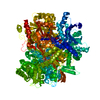
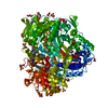
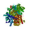
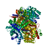
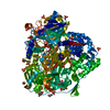
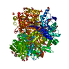
- PDB-4lgy: Importance of Hydrophobic Cavities in Allosteric Regulation of Fo... -
+ Open data

- Basic information
| Entry | Database: PDB / ID: 4lgy | ||||||
|---|---|---|---|---|---|---|---|
| Title | Importance of Hydrophobic Cavities in Allosteric Regulation of Formylglycinamide Synthetase: Insight from Xenon Trapping and Statistical Coupling Analysis | ||||||
 Components Components | Phosphoribosylformylglycinamidine synthase | ||||||
 Keywords Keywords | LIGASE / Amido transferase | ||||||
| Function / homology |  Function and homology informationphosphoribosylformylglycinamidine synthase / phosphoribosylformylglycinamidine synthase activity / purine nucleotide biosynthetic process / 'de novo' IMP biosynthetic process / glutamine metabolic process / ATP binding / metal ion binding / cytoplasm Function and homology informationphosphoribosylformylglycinamidine synthase / phosphoribosylformylglycinamidine synthase activity / purine nucleotide biosynthetic process / 'de novo' IMP biosynthetic process / glutamine metabolic process / ATP binding / metal ion binding / cytoplasmSimilarity search - Function | ||||||
| Biological species |  Salmonella typhimurium (bacteria) Salmonella typhimurium (bacteria) | ||||||
| Method | X-RAY DIFFRACTION / SYNCHROTRON / MOLECULAR REPLACEMENT / Resolution: 1.48 Å | ||||||
 Authors Authors | Tanwar, A.S. / Goyal, V.D. / Choudhary, D. / Panjikar, S. / Anand, R. | ||||||
 Citation Citation | Journal: Plos One / Year: 2013 Title: Importance of hydrophobic cavities in allosteric regulation of formylglycinamide synthetase: insight from xenon trapping and statistical coupling analysis Authors: Tanwar, A.S. / Goyal, V.D. / Choudhary, D. / Panjikar, S. / Anand, R. | ||||||
| History |
|
- Structure visualization
| Structure viewer | Molecule: MolmilJmol/JSmol |
|---|
- Downloads & links
-Download
| PDBx/mmCIF format | 4lgy.cif.gz | 320.7 KB | Display | PDBx/mmCIF format |
|---|---|---|---|---|
| PDB format | pdb4lgy.ent.gz | 245.7 KB | Display | PDB format |
| PDBx/mmJSON format | 4lgy.json.gz | Tree view | PDBx/mmJSON format | |
| Others |  Other downloads Other downloads |
-Validation report
| Arichive directory | https://data.pdbj.org/pub/pdb/validation_reports/lg/4lgyftp://data.pdbj.org/pub/pdb/validation_reports/lg/4lgy | HTTPS FTP |
|---|
-Related structure data
| Related structure data |  4l78C 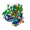 4mghC  1t3tS S: Starting model for refinement C: citing same article ( |
|---|---|
| Similar structure data |


-Links
 PDBj
PDBj


- Assembly
| Deposited unit | 
| ||||||||
|---|---|---|---|---|---|---|---|---|---|
| 1 |
| ||||||||
| Unit cell |
|
-Components
-Protein , 1 types, 1 molecules A
| #1: Protein | / FGAM synthase / FGAMS / Formylglycinamide ribotide amidotransferase / FGARAT / Formylglycinamide ...FGAM synthase / FGAMS / Formylglycinamide ribotide amidotransferase / FGARAT / Formylglycinamide ribotide synthetase Mass: 142777.594 Da / Num. of mol.: 1 / Mutation: F209W Source method: isolated from a genetically manipulated source Source: (gene. exp.) Salmonella typhimurium (bacteria) / Strain: LT2 / SGSC1412 / ATCC 700720 / Production host: Escherichia coli (E. coli)References: UniProt: P74881, phosphoribosylformylglycinamidine synthase |
|---|
-Non-polymers , 7 types, 1650 molecules 


| #2: Chemical | ChemComp-ADP / Adenosine diphosphate Mass: 427.201 Da / Num. of mol.: 1 / Source method: obtained synthetically / Formula: C10H15N5O10P2 / Comment: ADP, energy-carrying molecule*YM Mass: 427.201 Da / Num. of mol.: 1 / Source method: obtained synthetically / Formula: C10H15N5O10P2 / Comment: ADP, energy-carrying molecule*YM | ||||||||||
|---|---|---|---|---|---|---|---|---|---|---|---|
| #3: Chemical | ChemComp-MG /  Mass: 24.305 Da / Num. of mol.: 7 / Source method: obtained synthetically / Formula: Mg Mass: 24.305 Da / Num. of mol.: 7 / Source method: obtained synthetically / Formula: Mg#4: Chemical | ChemComp-SO4 / Sulfate Mass: 96.063 Da / Num. of mol.: 19 / Source method: obtained synthetically / Formula: SO4 Mass: 96.063 Da / Num. of mol.: 19 / Source method: obtained synthetically / Formula: SO4#5: Chemical | Acetate Mass: 59.044 Da / Num. of mol.: 3 / Source method: obtained synthetically / Formula: C2H3O2 Mass: 59.044 Da / Num. of mol.: 3 / Source method: obtained synthetically / Formula: C2H3O2#6: Chemical | ChemComp-GOL / Glycerol Mass: 92.094 Da / Num. of mol.: 4 / Source method: obtained synthetically / Formula: C3H8O3 Mass: 92.094 Da / Num. of mol.: 4 / Source method: obtained synthetically / Formula: C3H8O3#7: Chemical | ChemComp-CL / Chloride Mass: 35.453 Da / Num. of mol.: 6 / Source method: obtained synthetically / Formula: Cl Mass: 35.453 Da / Num. of mol.: 6 / Source method: obtained synthetically / Formula: Cl#8: Water | ChemComp-HOH / | WaterMass: 18.015 Da / Num. of mol.: 1610 / Source method: isolated from a natural source / Formula: H2O |
-Experimental details
-Experiment
| Experiment | Method: X-RAY DIFFRACTION / Number of used crystals: 1 |
|---|
- Sample preparation
| Crystal | Density Matthews: 3.05 Å3/Da / Density % sol: 59.65 % |
|---|---|
| Crystal grow | Method: vapor diffusion, hanging drop / pH: 7.5 Details: 2M ammonium sulphate, pH 7.5, VAPOR DIFFUSION, HANGING DROP |
-Data collection
| Diffraction source | Source: SYNCHROTRON / Site: ESRF  / Beamline: BM14 / Wavelength: 0.97372 Å / Beamline: BM14 / Wavelength: 0.97372 Å |
|---|---|
| Detector | Type: MARMOSAIC 225 mm CCD / Detector: CCD / Date: Feb 13, 2013 |
| Radiation | Protocol: SINGLE WAVELENGTH / Monochromatic (M) / Laue (L): M / Scattering type: x-ray |
| Radiation wavelength | Wavelength: 0.97372 Å / Relative weight: 1 |
| Reflection | Resolution: 1.48→27.5 Å / Num. all: 274143 / Num. obs: 274143 / % possible obs: 96.5 % / Redundancy: 3.9 % / Rmerge(I) obs: 0.067 / Net I/σ(I): 11.3 |
| Reflection shell | Highest resolution: 1.48 Å |
- Processing
| Software |
| ||||||||||||||||||||||||||||||||||||||||||||||||||||||||||||||||||||||||||||||||||||||||||||||||||||||||||||||||||||||||||||||||||||||||||||||||||||||||||||||||||||||||||||||||||||||
|---|---|---|---|---|---|---|---|---|---|---|---|---|---|---|---|---|---|---|---|---|---|---|---|---|---|---|---|---|---|---|---|---|---|---|---|---|---|---|---|---|---|---|---|---|---|---|---|---|---|---|---|---|---|---|---|---|---|---|---|---|---|---|---|---|---|---|---|---|---|---|---|---|---|---|---|---|---|---|---|---|---|---|---|---|---|---|---|---|---|---|---|---|---|---|---|---|---|---|---|---|---|---|---|---|---|---|---|---|---|---|---|---|---|---|---|---|---|---|---|---|---|---|---|---|---|---|---|---|---|---|---|---|---|---|---|---|---|---|---|---|---|---|---|---|---|---|---|---|---|---|---|---|---|---|---|---|---|---|---|---|---|---|---|---|---|---|---|---|---|---|---|---|---|---|---|---|---|---|---|---|---|---|---|
| Refinement | Method to determine structure: MOLECULAR REPLACEMENT Starting model: 1T3T Resolution: 1.48→27.5 Å / Cor.coef. Fo:Fc: 0.971 / Cor.coef. Fo:Fc free: 0.962 / Occupancy max: 1 / Occupancy min: 0.25 / SU B: 0.749 / SU ML: 0.029 / Cross valid method: THROUGHOUT / ESU R: 0.049 / ESU R Free: 0.052 / Stereochemistry target values: MAXIMUM LIKELIHOOD / Details: HYDROGENS HAVE BEEN USED IF PRESENT IN THE INPUT
| ||||||||||||||||||||||||||||||||||||||||||||||||||||||||||||||||||||||||||||||||||||||||||||||||||||||||||||||||||||||||||||||||||||||||||||||||||||||||||||||||||||||||||||||||||||||
| Solvent computation | Ion probe radii: 0.8 Å / Shrinkage radii: 0.8 Å / VDW probe radii: 1.2 Å / Solvent model: BABINET MODEL WITH MASK | ||||||||||||||||||||||||||||||||||||||||||||||||||||||||||||||||||||||||||||||||||||||||||||||||||||||||||||||||||||||||||||||||||||||||||||||||||||||||||||||||||||||||||||||||||||||
| Displacement parameters | Biso mean: 14.885 Å2
| ||||||||||||||||||||||||||||||||||||||||||||||||||||||||||||||||||||||||||||||||||||||||||||||||||||||||||||||||||||||||||||||||||||||||||||||||||||||||||||||||||||||||||||||||||||||
| Refinement step | Cycle: LAST / Resolution: 1.48→27.5 Å
| ||||||||||||||||||||||||||||||||||||||||||||||||||||||||||||||||||||||||||||||||||||||||||||||||||||||||||||||||||||||||||||||||||||||||||||||||||||||||||||||||||||||||||||||||||||||
| Refine LS restraints |
|