[English] 日本語
Yorodumi
- PDB-4lab: Crystal structure of the catalytic domain of RluB -

+
Open data


ID or keywords:

Loading...

-
Basic information

Entry
Database: PDB / ID: 4lab
TitleCrystal structure of the catalytic domain of RluB
ComponentsRibosomal large subunit pseudouridine synthase B
KeywordsRNA BINDING PROTEIN / beta sheet / alpha-beta protein / pseudouridine synthase / E. coli ribosomal RNA
Function / homology
Function and homology information


23S rRNA pseudouridine2605 synthase / rRNA pseudouridine synthase activity / enzyme-directed rRNA pseudouridine synthesis / RNA binding / cytosol
Similarity search - Function
Pseudouridine synthase, RsuA/RluB/E/F / Pseudouridine synthase, RsuA/RluB/E/F, conserved site / Rsu family of pseudouridine synthase signature. / Pseudouridine synthase, RsuA/RluA-like / RNA pseudouridylate synthase / Pseudouridine synthase / Pseudouridine synthase / Pseudouridine synthase, catalytic domain superfamily / S4 RNA-binding domain / RNA-binding S4 domain ...Pseudouridine synthase, RsuA/RluB/E/F / Pseudouridine synthase, RsuA/RluB/E/F, conserved site / Rsu family of pseudouridine synthase signature. / Pseudouridine synthase, RsuA/RluA-like / RNA pseudouridylate synthase / Pseudouridine synthase / Pseudouridine synthase / Pseudouridine synthase, catalytic domain superfamily / S4 RNA-binding domain / RNA-binding S4 domain / RNA-binding S4 domain superfamily / S4 domain / S4 RNA-binding domain profile. / 2-Layer Sandwich / Alpha Beta
Similarity search - Domain/homology
: / Ribosomal large subunit pseudouridine synthase B
Similarity search - Component
Biological speciesEscherichia coli (E. coli)
MethodX-RAY DIFFRACTION / SYNCHROTRON / MOLECULAR REPLACEMENT / Resolution: 2.5043 Å
AuthorsCzudnochowski, N. / Finer-Moore, J.S. / Stroud, R.M.
CitationJournal: Nucleic Acids Res. / Year: 2014
Title: The mechanism of pseudouridine synthases from a covalent complex with RNA, and alternate specificity for U2605 versus U2604 between close homologs.
Authors: Czudnochowski, N. / Ashley, G.W. / Santi, D.V. / Alian, A. / Finer-Moore, J. / Stroud, R.M.
History
DepositionJun 19, 2013Deposition site: RCSB / Processing site: RCSB
Revision 1.0Nov 20, 2013Provider: repository / Type: Initial release
Revision 1.1Feb 26, 2014Group: Database references
Revision 1.2Nov 15, 2017Group: Refinement description / Category: software / Item: _software.classification
Revision 1.3Jul 17, 2019Group: Data collection / Refinement description / Category: software / Item: _software.name / _software.version
Revision 1.4Sep 20, 2023Group: Data collection / Database references ...Data collection / Database references / Derived calculations / Refinement description
Category: chem_comp_atom / chem_comp_bond ...chem_comp_atom / chem_comp_bond / database_2 / pdbx_initial_refinement_model / pdbx_struct_conn_angle / struct_conn / struct_ref_seq_dif / struct_site
Item: _database_2.pdbx_DOI / _database_2.pdbx_database_accession ..._database_2.pdbx_DOI / _database_2.pdbx_database_accession / _pdbx_struct_conn_angle.ptnr1_auth_comp_id / _pdbx_struct_conn_angle.ptnr1_auth_seq_id / _pdbx_struct_conn_angle.ptnr1_label_atom_id / _pdbx_struct_conn_angle.ptnr1_label_comp_id / _pdbx_struct_conn_angle.ptnr1_label_seq_id / _pdbx_struct_conn_angle.ptnr3_auth_comp_id / _pdbx_struct_conn_angle.ptnr3_auth_seq_id / _pdbx_struct_conn_angle.ptnr3_label_atom_id / _pdbx_struct_conn_angle.ptnr3_label_comp_id / _pdbx_struct_conn_angle.ptnr3_label_seq_id / _pdbx_struct_conn_angle.value / _struct_conn.pdbx_dist_value / _struct_conn.ptnr1_auth_comp_id / _struct_conn.ptnr1_auth_seq_id / _struct_conn.ptnr1_label_atom_id / _struct_conn.ptnr1_label_comp_id / _struct_conn.ptnr1_label_seq_id / _struct_conn.ptnr2_auth_seq_id / _struct_conn.ptnr2_label_asym_id / _struct_ref_seq_dif.details / _struct_site.pdbx_auth_asym_id / _struct_site.pdbx_auth_comp_id / _struct_site.pdbx_auth_seq_id

-
Structure visualization

Structure viewerMolecule:
MolmilJmol/JSmol

Downloads & links

-
Assembly

Deposited unit
A: Ribosomal large subunit pseudouridine synthase B
hetero molecules


Theoretical massNumber of molelcules
Total (without water)28,9784
Polymers28,5531
Non-polymers4263
Water34219
1


  • Idetical with deposited unit
  • defined by author&software
TypeNameSymmetry operationNumber
identity operation1_555x,y,z1
MethodPISA
Unit cell
Length a, b, c (Å)82.390, 82.390, 74.900
Angle α, β, γ (deg.)90.00, 90.00, 90.00
Int Tables number96
Space group name H-MP43212

-
Components

#1: Protein Ribosomal large subunit pseudouridine synthase B / / 23S rRNA pseudouridine(2605) synthase / rRNA pseudouridylate synthase B / rRNA-uridine isomerase B


Mass: 28552.619 Da / Num. of mol.: 1 / Fragment: UNP Residues 1-251
Source method: isolated from a genetically manipulated source
Source: (gene. exp.) Escherichia coli (E. coli) / Strain: K12 / Gene: b1269, JW1261, rluB, yciL / Plasmid: pET47 modified / Production host: Escherichia coli (E. coli) / Strain (production host): BL21(DE3)
References: UniProt: P37765, 23S rRNA pseudouridine2605 synthase
#2: Chemical ChemComp-PT / PLATINUM (II) ION


Mass: 195.078 Da / Num. of mol.: 2 / Source method: obtained synthetically / Formula: Pt
#3: Chemical ChemComp-CL / CHLORIDE ION / Chloride


Mass: 35.453 Da / Num. of mol.: 1 / Source method: obtained synthetically / Formula: Cl
#4: Water ChemComp-HOH / water / Water


Mass: 18.015 Da / Num. of mol.: 19 / Source method: isolated from a natural source / Formula: H2O

-
Experimental details

-
Experiment

ExperimentMethod: X-RAY DIFFRACTION / Number of used crystals: 1

-
Sample preparation

CrystalDensity Matthews: 2.23 Å3/Da / Density % sol: 44.74 %
Crystal growTemperature: 289 K / Method: vapor diffusion, hanging drop / pH: 5.6
Details: 0.1M tri-Sodium citrate pH 5.6, 20% isopropanol, 15% PEG4000, VAPOR DIFFUSION, HANGING DROP, temperature 289K

-
Data collection

DiffractionMean temperature: 100 K
Diffraction sourceSource: SYNCHROTRON / Site: ALS / Beamline: 8.3.1 / Wavelength: 1.06883 Å
DetectorType: ADSC QUANTUM 315r / Detector: CCD / Date: Feb 17, 2011 / Details: monochromator
RadiationMonochromator: Si(111) / Protocol: SINGLE WAVELENGTH / Monochromatic (M) / Laue (L): M / Scattering type: x-ray
Radiation wavelengthWavelength: 1.06883 Å / Relative weight: 1
ReflectionResolution: 2.5→58.259 Å / Num. all: 9324 / Num. obs: 9324 / % possible obs: 99.4 % / Observed criterion σ(I): -3 / Redundancy: 18.9 % / Biso Wilson estimate: 56.5 Å2 / Rmerge(I) obs: 0.099 / Net I/σ(I): 21.52
Reflection shellResolution: 2.5→2.6 Å / Redundancy: 17.5 % / Rmerge(I) obs: 1.53 / Mean I/σ(I) obs: 2.6 / % possible all: 94.2

-
Processing

Software
NameVersionClassification
PHENIX(phenix.refine: 1.8_1069)refinement
PHENIXmodel building
ELVESrefinement
XDSdata reduction
XDSdata scaling
PHENIXphasing
RefinementMethod to determine structure: MOLECULAR REPLACEMENT
Starting model: PDB ENTRY 2GML
Resolution: 2.5043→58.259 Å / SU ML: 0.3
Isotropic thermal model: Isotropic with TLS parameters for 2 groups
σ(F): 2 / Phase error: 31.41 / Stereochemistry target values: ML
RfactorNum. reflection% reflection
Rfree0.2605 471 5.06 %
Rwork0.2306 --
obs0.2322 9317 99.82 %
all-9317 -
Solvent computationShrinkage radii: 1.2 Å / VDW probe radii: 1.3 Å / Solvent model: FLAT BULK SOLVENT MODEL
Refinement stepCycle: LAST / Resolution: 2.5043→58.259 Å
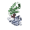
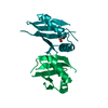
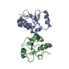
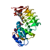
ProteinNucleic acidLigandSolventTotal
Num. atoms1459 0 3 19 1481
Refine LS restraints
Refine-IDTypeDev idealNumber
X-RAY DIFFRACTIONf_bond_d0.0061494
X-RAY DIFFRACTIONf_angle_d0.9172029
X-RAY DIFFRACTIONf_dihedral_angle_d14.75553
X-RAY DIFFRACTIONf_chiral_restr0.06225
X-RAY DIFFRACTIONf_plane_restr0.005268
LS refinement shell
Resolution (Å)Rfactor RfreeNum. reflection RfreeRfactor RworkNum. reflection RworkRefine-ID% reflection obs (%)
2.5043-2.86660.31981480.27782867X-RAY DIFFRACTION100
2.8666-3.61160.29741470.25442921X-RAY DIFFRACTION100
3.6116-58.27430.23621760.21233058X-RAY DIFFRACTION100
Refinement TLS params.

Method: refined / Refine-ID: X-RAY DIFFRACTION

IDL112)L122)L132)L222)L232)L332)S11 (Å °)S12 (Å °)S13 (Å °)S21 (Å °)S22 (Å °)S23 (Å °)S31 (Å °)S32 (Å °)S33 (Å °)T112)T122)T132)T222)T232)T332)Origin x (Å)Origin y (Å)Origin z (Å)
13.92932.7754-0.08738.1207-0.43514.896-0.07550.47650.095-0.46160.0403-0.3759-0.02290.4250.07180.26720.05260.00070.49860.04230.280640.88838.897924.98
21.09670.5513-0.74874.9251-3.02064.48860.16680.17740.35920.42640.21780.6725-0.6925-0.6268-0.33110.37870.07610.10620.54220.10860.580334.375823.670627.2208
Refinement TLS group
IDRefine-IDRefine TLS-IDSelection details
1X-RAY DIFFRACTION1(chain A and resid 65:138)
2X-RAY DIFFRACTION2(chain A and resid 139:251)

+
About Yorodumi

-
News

-
Feb 9, 2022. New format data for meta-information of EMDB entries

New format data for meta-information of EMDB entries

  • Version 3 of the EMDB header file is now the official format.
  • The previous official version 1.9 will be removed from the archive.

Related info.:EMDB header

External links:wwPDB to switch to version 3 of the EMDB data model

-
Aug 12, 2020. Covid-19 info

Covid-19 info

URL: https://pdbj.org/emnavi/covid19.php

New page: Covid-19 featured information page in EM Navigator.

Related info.:Covid-19 info / Mar 5, 2020. Novel coronavirus structure data

+
Mar 5, 2020. Novel coronavirus structure data

Novel coronavirus structure data

Related info.:Yorodumi Speices / Aug 12, 2020. Covid-19 info

External links:COVID-19 featured content - PDBj / Molecule of the Month (242):Coronavirus Proteases

+
Jan 31, 2019. EMDB accession codes are about to change! (news from PDBe EMDB page)

EMDB accession codes are about to change! (news from PDBe EMDB page)

  • The allocation of 4 digits for EMDB accession codes will soon come to an end. Whilst these codes will remain in use, new EMDB accession codes will include an additional digit and will expand incrementally as the available range of codes is exhausted. The current 4-digit format prefixed with “EMD-” (i.e. EMD-XXXX) will advance to a 5-digit format (i.e. EMD-XXXXX), and so on. It is currently estimated that the 4-digit codes will be depleted around Spring 2019, at which point the 5-digit format will come into force.
  • The EM Navigator/Yorodumi systems omit the EMD- prefix.

Related info.:Q: What is EMD? / ID/Accession-code notation in Yorodumi/EM Navigator

External links:EMDB Accession Codes are Changing Soon! / Contact to PDBj

+
Jul 12, 2017. Major update of PDB

Major update of PDB

  • wwPDB released updated PDB data conforming to the new PDBx/mmCIF dictionary.
  • This is a major update changing the version number from 4 to 5, and with Remediation, in which all the entries are updated.
  • In this update, many items about electron microscopy experimental information are reorganized (e.g. em_software).
  • Now, EM Navigator and Yorodumi are based on the updated data.

External links:wwPDB Remediation / Enriched Model Files Conforming to OneDep Data Standards Now Available in the PDB FTP Archive

-
Yorodumi

Thousand views of thousand structures

  • Yorodumi is a browser for structure data from EMDB, PDB, SASBDB, etc.
  • This page is also the successor to EM Navigator detail page, and also detail information page/front-end page for Omokage search.
  • The word "yorodu" (or yorozu) is an old Japanese word meaning "ten thousand". "mi" (miru) is to see.

Related info.:EMDB / PDB / SASBDB / Comparison of 3 databanks / Yorodumi Search / Aug 31, 2016. New EM Navigator & Yorodumi / Yorodumi Papers / Jmol/JSmol / Function and homology information / Changes in new EM Navigator and Yorodumi

Read more