 Movie
Movie Controller


+ Open data

- Basic information
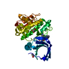
| Entry | Database: PDB / ID: 4l3j | ||||||
|---|---|---|---|---|---|---|---|
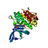
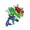
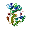
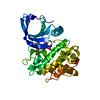
| Title | Crystal structures of human p70S6K1 kinase domain | ||||||
 Components Components | RPS6KB1 protein P70-S6 Kinase 1 P70-S6 Kinase 1 | ||||||
 Keywords Keywords | TRANSFERASE / Protein kinase | ||||||
| Function / homology |  Function and homology information Function and homology informationlong-chain fatty acid import into cell / response to electrical stimulus involved in regulation of muscle adaptation / skeletal muscle atrophy / positive regulation of skeletal muscle tissue growth / regulation of glucose import / ribosomal protein S6 kinase activity / response to L-leucine / cellular response to nutrient / phosphatidylinositol-mediated signaling / response to glucagon ...long-chain fatty acid import into cell / response to electrical stimulus involved in regulation of muscle adaptation / skeletal muscle atrophy / positive regulation of skeletal muscle tissue growth / regulation of glucose import / ribosomal protein S6 kinase activity / response to L-leucine / cellular response to nutrient / phosphatidylinositol-mediated signaling / response to glucagon / response to testosterone / positive regulation of smooth muscle cell migration / TOR signaling / mTORC1-mediated signalling / germ cell development / positive regulation of translational initiation / skeletal muscle contraction / long-term memory / behavioral fear response / response to tumor necrosis factor / response to glucose / response to mechanical stimulus / negative regulation of insulin receptor signaling pathway / positive regulation of TORC1 signaling / protein serine/threonine/tyrosine kinase activity / cellular response to dexamethasone stimulus / positive regulation of mitotic cell cycle / response to nutrient levels / protein phosphatase 2A binding / positive regulation of translation / phosphatidylinositol 3-kinase/protein kinase B signal transduction / PDZ domain binding / negative regulation of extrinsic apoptotic signaling pathway / peptide binding / positive regulation of smooth muscle cell proliferation / G1/S transition of mitotic cell cycle / modulation of chemical synaptic transmission / response to toxic substance / cellular response to growth factor stimulus / cellular response to type II interferon / cellular response to insulin stimulus / cell migration / postsynapse / peptidyl-serine phosphorylation / response to ethanol / mitochondrial outer membrane / response to lipopolysaccharide / non-specific serine/threonine protein kinase / neuron projection / protein kinase activity / response to xenobiotic stimulus / protein serine kinase activity / protein serine/threonine kinase activity / glutamatergic synapse / apoptotic process / negative regulation of apoptotic process / perinuclear region of cytoplasm / cell surface / signal transduction / mitochondrion / nucleoplasm / ATP binding / identical protein binding / nucleus / cytosol / cytoplasmSimilarity search - Function | ||||||
| Biological species |  Homo sapiens (human) Homo sapiens (human) | ||||||
| Method | X-RAY DIFFRACTION / SYNCHROTRON / MOLECULAR REPLACEMENT / Resolution: 2.1 Å | ||||||
 Authors Authors | Wang, J. / Zhong, C. / Ding, J. | ||||||
 Citation Citation | Journal: Biochem.J. / Year: 2013 Title: Crystal structures of S6K1 provide insights into the regulation mechanism of S6K1 by the hydrophobic motif Authors: Wang, J. / Zhong, C. / Wang, F. / Qu, F. / Ding, J. | ||||||
| History |
|
- Structure visualization
| Structure viewer | Molecule: MolmilJmol/JSmol |
|---|
- Downloads & links
-Download
| PDBx/mmCIF format | 4l3j.cif.gz | 138.2 KB | Display | PDBx/mmCIF format |
|---|---|---|---|---|
| PDB format | pdb4l3j.ent.gz | 107.4 KB | Display | PDB format |
| PDBx/mmJSON format | 4l3j.json.gz | Tree view | PDBx/mmJSON format | |
| Others |  Other downloads Other downloads |
-Validation report
| Arichive directory | https://data.pdbj.org/pub/pdb/validation_reports/l3/4l3jftp://data.pdbj.org/pub/pdb/validation_reports/l3/4l3j | HTTPS FTP |
|---|
-Related structure data
| Related structure data | 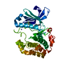 4l3lC 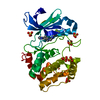 4l42C 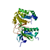 4l43C 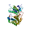 4l44C 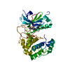 4l45C 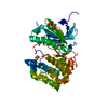 4l46C 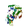 3a62S C: citing same article ( S: Starting model for refinement |
|---|---|
| Similar structure data |


-Links
 PDBj
PDBj


- Assembly
| Deposited unit | 
| ||||||||
|---|---|---|---|---|---|---|---|---|---|
| 1 |
| ||||||||
| Unit cell |
|
-Components
| #1: Protein | P70-S6 Kinase 1 / Ribosomal protein S6 kinase beta-1 / Ribosomal protein S6 kinase / 70kDa / polypeptide 1 / isoform CRA_c Mass: 34337.094 Da / Num. of mol.: 1 Source method: isolated from a genetically manipulated source Source: (gene. exp.) Homo sapiens (human) / Gene: RPS6KB1, hCG_1815774 / Plasmid: pFastBacHTB / Production host:   Spodoptera frugiperda (fall armyworm) / Strain (production host): Spodoptera frugiperda / References: UniProt: Q7Z721, UniProt: P23443*PLUS Spodoptera frugiperda (fall armyworm) / Strain (production host): Spodoptera frugiperda / References: UniProt: Q7Z721, UniProt: P23443*PLUS |
|---|---|
| #2: Chemical | ChemComp-5FI /   Mass: 390.405 Da / Num. of mol.: 1 / Source method: obtained synthetically / Formula: C19H21F3N6 Mass: 390.405 Da / Num. of mol.: 1 / Source method: obtained synthetically / Formula: C19H21F3N6 |
| #3: Chemical | ChemComp-ZN /   Mass: 65.409 Da / Num. of mol.: 1 / Source method: obtained synthetically / Formula: Zn Mass: 65.409 Da / Num. of mol.: 1 / Source method: obtained synthetically / Formula: Zn |
| #4: Water | ChemComp-HOH / Water Mass: 18.015 Da / Num. of mol.: 121 / Source method: isolated from a natural source / Formula: H2O Mass: 18.015 Da / Num. of mol.: 121 / Source method: isolated from a natural source / Formula: H2O |
-Experimental details
-Experiment
| Experiment | Method: X-RAY DIFFRACTION / Number of used crystals: 1 |
|---|
- Sample preparation
| Crystal | Density Matthews: 2.82 Å3/Da / Density % sol: 56.34 % |
|---|
-Data collection
| Diffraction | Mean temperature: 100 K |
|---|---|
| Diffraction source | Source: SYNCHROTRON / Site: SSRF  / Beamline: BL17U / Wavelength: 1 Å / Beamline: BL17U / Wavelength: 1 Å |
| Detector | Type: CCD ADSC unsupported-q315 / Detector: CCD / Date: Jun 20, 2011 |
| Radiation | Protocol: SINGLE WAVELENGTH / Monochromatic (M) / Laue (L): M / Scattering type: x-ray |
| Radiation wavelength | Wavelength: 1 Å / Relative weight: 1 |
| Reflection | Resolution: 2.1→50 Å / Num. obs: 22617 |
- Processing
| Software |
| ||||||||||||||||||||||||||||||||||||||||||||||||||||||||||||||||||||||
|---|---|---|---|---|---|---|---|---|---|---|---|---|---|---|---|---|---|---|---|---|---|---|---|---|---|---|---|---|---|---|---|---|---|---|---|---|---|---|---|---|---|---|---|---|---|---|---|---|---|---|---|---|---|---|---|---|---|---|---|---|---|---|---|---|---|---|---|---|---|---|---|
| Refinement | Method to determine structure: MOLECULAR REPLACEMENT Starting model: 3A62 Resolution: 2.1→50 Å / Cor.coef. Fo:Fc: 0.953 / Cor.coef. Fo:Fc free: 0.934 / Occupancy max: 1 / Occupancy min: 0.57 / SU B: 14.348 / SU ML: 0.162 / Cross valid method: THROUGHOUT / σ(F): 0 / ESU R Free: 0.187 / Stereochemistry target values: MAXIMUM LIKELIHOOD / Details: U VALUES: RESIDUAL ONLY
| ||||||||||||||||||||||||||||||||||||||||||||||||||||||||||||||||||||||
| Solvent computation | Ion probe radii: 0.8 Å / Shrinkage radii: 0.8 Å / VDW probe radii: 1.4 Å / Solvent model: MASK | ||||||||||||||||||||||||||||||||||||||||||||||||||||||||||||||||||||||
| Displacement parameters | Biso max: 129.48 Å2 / Biso mean: 47.2591 Å2 / Biso min: 18.06 Å2
| ||||||||||||||||||||||||||||||||||||||||||||||||||||||||||||||||||||||
| Refinement step | Cycle: LAST / Resolution: 2.1→50 Å
| ||||||||||||||||||||||||||||||||||||||||||||||||||||||||||||||||||||||
| Refine LS restraints |
| ||||||||||||||||||||||||||||||||||||||||||||||||||||||||||||||||||||||
| LS refinement shell | Resolution: 2.1→2.155 Å / Total num. of bins used: 20
| ||||||||||||||||||||||||||||||||||||||||||||||||||||||||||||||||||||||
| Refinement TLS params. | Method: refined / Origin x: 23.2796 Å / Origin y: -13.423 Å / Origin z: 6.0683 Å
|
