 Movie
Movie Controller

[English] 日本語
 Yorodumi
- PDB-4kon: The structure of hemagglutinin from avian-origin H7N9 influenza v... -
+ Open data

- Basic information
| Entry | Database: PDB / ID: 4kon | |||||||||
|---|---|---|---|---|---|---|---|---|---|---|
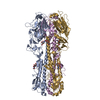
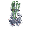
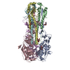
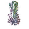
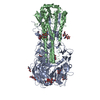
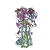
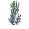
| Title | The structure of hemagglutinin from avian-origin H7N9 influenza virus in complex with human receptor analog 6'SLNLN (NeuAcα2-6Galβ1-4GlcNAcβ1-3Galβ1-4Glc) | |||||||||
 Components Components |
| |||||||||
 Keywords Keywords |  VIRAL PROTEIN / homotrimer / virus attachment and membrane fusion VIRAL PROTEIN / homotrimer / virus attachment and membrane fusion | |||||||||
| Function / homology |  Function and homology information Function and homology informationclathrin-dependent endocytosis of virus by host cell / host cell surface receptor binding / apical plasma membrane / fusion of virus membrane with host plasma membrane / fusion of virus membrane with host endosome membrane / viral envelope / virion attachment to host cell / host cell plasma membraneSimilarity search - Function | |||||||||
| Biological species |   Influenza A virus Influenza A virus | |||||||||
| Method | X-RAY DIFFRACTION / SYNCHROTRON / MOLECULAR REPLACEMENT / Resolution: 2.6 Å | |||||||||
 Authors Authors | Shi, Y. / Zhang, W. / Wang, F. / Qi, J. / Song, H. / Wu, Y. / Gao, F. / Zhang, Y. / Fan, Z. / Gong, W. ...Shi, Y. / Zhang, W. / Wang, F. / Qi, J. / Song, H. / Wu, Y. / Gao, F. / Zhang, Y. / Fan, Z. / Gong, W. / Wang, D. / Shu, Y. / Wang, Y. / Yan, J. / Gao, G.F. | |||||||||
 Citation Citation | Journal: Science / Year: 2013 Title: Structures and receptor binding of hemagglutinins from human-infecting H7N9 influenza viruses. Authors: Shi, Y. / Zhang, W. / Wang, F. / Qi, J. / Wu, Y. / Song, H. / Gao, F. / Bi, Y. / Zhang, Y. / Fan, Z. / Qin, C. / Sun, H. / Liu, J. / Haywood, J. / Liu, W. / Gong, W. / Wang, D. / Shu, Y. / ...Authors: Shi, Y. / Zhang, W. / Wang, F. / Qi, J. / Wu, Y. / Song, H. / Gao, F. / Bi, Y. / Zhang, Y. / Fan, Z. / Qin, C. / Sun, H. / Liu, J. / Haywood, J. / Liu, W. / Gong, W. / Wang, D. / Shu, Y. / Wang, Y. / Yan, J. / Gao, G.F. | |||||||||
| History |
|
- Structure visualization
| Structure viewer | Molecule: MolmilJmol/JSmol |
|---|
- Downloads & links
-Download
| PDBx/mmCIF format | 4kon.cif.gz | 206.4 KB | Display | PDBx/mmCIF format |
|---|---|---|---|---|
| PDB format | pdb4kon.ent.gz | 168.8 KB | Display | PDB format |
| PDBx/mmJSON format | 4kon.json.gz | Tree view | PDBx/mmJSON format | |
| Others |  Other downloads Other downloads |
-Validation report
| Arichive directory | https://data.pdbj.org/pub/pdb/validation_reports/ko/4konftp://data.pdbj.org/pub/pdb/validation_reports/ko/4kon | HTTPS FTP |
|---|
-Related structure data
| Related structure data | 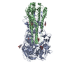 4kolC  4komC  4lcxC  4lkgC  4lkhC  4lkiC  4lkjC  4lkkC C: citing same article ( |
|---|---|
| Similar structure data |


-Links
 PDBj
PDBj


- Assembly
| Deposited unit | 
| ||||||||||||
|---|---|---|---|---|---|---|---|---|---|---|---|---|---|
| 1 | 
| ||||||||||||
| Unit cell |
| ||||||||||||
| Components on special symmetry positions |
|
-Components
| #1: Protein | Mass: 34195.594 Da / Num. of mol.: 1 Source method: isolated from a genetically manipulated source Source: (gene. exp.) Influenza A virus / Strain: A/Anhui/1/2013 (H7N9) / Gene: HA / Plasmid: pFastbac1 / Production host:  Trichoplusia ni (cabbage looper) / References: UniProt: V5IRU4*PLUS Trichoplusia ni (cabbage looper) / References: UniProt: V5IRU4*PLUS | ||
|---|---|---|---|
| #2: Protein | Mass: 19490.309 Da / Num. of mol.: 1 Source method: isolated from a genetically manipulated source Source: (gene. exp.) Influenza A virus / Strain: A/Anhui/1/2013 (H7N9) / Gene: HA / Plasmid: pFastbac1 / Production host: Trichoplusia ni (cabbage looper) / References: UniProt: V5IRU3*PLUS | ||
| #3: Polysaccharide | N-acetyl-alpha-neuraminic acid-(2-6)-beta-D-galactopyranose / Mass: 471.411 Da / Num. of mol.: 1 Source method: isolated from a genetically manipulated source | ||
| #4: Sugar | N-Acetylglucosamine  Type: D-saccharide, beta linking / Mass: 221.208 Da / Num. of mol.: 3 Type: D-saccharide, beta linking / Mass: 221.208 Da / Num. of mol.: 3Source method: isolated from a genetically manipulated source Formula: C8H15NO6 #5: Water | ChemComp-HOH / | Water Mass: 18.015 Da / Num. of mol.: 88 / Source method: isolated from a natural source / Formula: H2O Mass: 18.015 Da / Num. of mol.: 88 / Source method: isolated from a natural source / Formula: H2O |
-Experimental details
-Experiment
| Experiment | Method: X-RAY DIFFRACTION / Number of used crystals: 1 |
|---|
- Sample preparation
| Crystal | Density Matthews: 3.58 Å3/Da / Density % sol: 65.69 % |
|---|---|
| Crystal grow | Temperature: 291 K / Method: vapor diffusion, sitting drop / pH: 8.5 Details: 16% w/v PEG 3350, 0.2 M lithium sulfate, 0.1 M Tris, pH 8.5, VAPOR DIFFUSION, SITTING DROP, temperature 291K |
-Data collection
| Diffraction | Mean temperature: 100 K |
|---|---|
| Diffraction source | Source: SYNCHROTRON / Site: SSRF  / Beamline: BL17U / Wavelength: 1 Å / Beamline: BL17U / Wavelength: 1 Å |
| Detector | Type: ADSC QUANTUM 315r / Detector: CCD / Date: Apr 30, 2013 |
| Radiation | Monochromator: GRAPHITE / Protocol: SINGLE WAVELENGTH / Monochromatic (M) / Laue (L): M / Scattering type: x-ray |
| Radiation wavelength | Wavelength: 1 Å / Relative weight: 1 |
| Reflection | Resolution: 2.6→50 Å / Num. obs: 24064 / % possible obs: 99.9 % / Observed criterion σ(F): 0 / Observed criterion σ(I): -3 |
| Reflection shell | Resolution: 2.6→2.69 Å / % possible all: 100 |
- Processing
| Software |
| ||||||||||||||||||||||||||||||||||||||||||||||||||||||||||||||||||||||
|---|---|---|---|---|---|---|---|---|---|---|---|---|---|---|---|---|---|---|---|---|---|---|---|---|---|---|---|---|---|---|---|---|---|---|---|---|---|---|---|---|---|---|---|---|---|---|---|---|---|---|---|---|---|---|---|---|---|---|---|---|---|---|---|---|---|---|---|---|---|---|---|
| Refinement | Method to determine structure: MOLECULAR REPLACEMENT / Resolution: 2.6→47.654 Å / SU ML: 0.36 / σ(F): 1.35 / Phase error: 25.87 / Stereochemistry target values: ML
| ||||||||||||||||||||||||||||||||||||||||||||||||||||||||||||||||||||||
| Solvent computation | Shrinkage radii: 0.98 Å / VDW probe radii: 1.2 Å / Solvent model: FLAT BULK SOLVENT MODEL / Bsol: 35.038 Å2 / ksol: 0.315 e/Å3 | ||||||||||||||||||||||||||||||||||||||||||||||||||||||||||||||||||||||
| Displacement parameters |
| ||||||||||||||||||||||||||||||||||||||||||||||||||||||||||||||||||||||
| Refinement step | Cycle: LAST / Resolution: 2.6→47.654 Å
| ||||||||||||||||||||||||||||||||||||||||||||||||||||||||||||||||||||||
| Refine LS restraints |
| ||||||||||||||||||||||||||||||||||||||||||||||||||||||||||||||||||||||
| LS refinement shell |
| ||||||||||||||||||||||||||||||||||||||||||||||||||||||||||||||||||||||
| Refinement TLS params. | Method: refined / Origin x: -15.7678 Å / Origin y: -3.8179 Å / Origin z: -56.9091 Å
| ||||||||||||||||||||||||||||||||||||||||||||||||||||||||||||||||||||||
| Refinement TLS group | Selection details: all |
