 Movie
Movie Controller

[English] 日本語
 Yorodumi
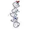
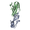
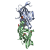
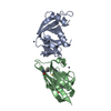
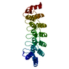
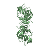
- PDB-4jmh: Crystal structure of synthetic protein in complex with double pY ... -
+ Open data

- Basic information
| Entry | Database: PDB / ID: 4jmh | ||||||
|---|---|---|---|---|---|---|---|
| Title | Crystal structure of synthetic protein in complex with double pY peptide | ||||||
 Components Components |
| ||||||
 Keywords Keywords |  DE NOVO PROTEIN / Synthetic protein / binding to double pY containing sequence DE NOVO PROTEIN / Synthetic protein / binding to double pY containing sequence | ||||||
| Function / homology |  Function and homology information Function and homology informationregulation of superoxide metabolic process / positive regulation of cell proliferation in bone marrow / XBP1(S) activates chaperone genes / neurotrophin TRKA receptor binding / transmembrane receptor protein tyrosine kinase adaptor activity / Interleukin-15 signaling / Shc-EGFR complex / Interleukin-2 signaling / epidermal growth factor binding / epidermal growth factor receptor binding ...regulation of superoxide metabolic process / positive regulation of cell proliferation in bone marrow / XBP1(S) activates chaperone genes / neurotrophin TRKA receptor binding / transmembrane receptor protein tyrosine kinase adaptor activity / Interleukin-15 signaling / Shc-EGFR complex / Interleukin-2 signaling / epidermal growth factor binding / epidermal growth factor receptor binding / Signaling by ALK / Signaling by ALK fusions and activated point mutants / Activated NTRK3 signals through RAS / RET signaling / Activated NTRK2 signals through RAS / Interleukin-3, Interleukin-5 and GM-CSF signaling / SHC1 events in ERBB4 signaling / Signalling to RAS / SHC-related events triggered by IGF1R / Role of LAT2/NTAL/LAB on calcium mobilization / Signal attenuation / Interleukin receptor SHC signaling / SHC-mediated cascade:FGFR3 / MET activates RAS signaling / SHC-mediated cascade:FGFR2 / SHC-mediated cascade:FGFR4 / SHC-mediated cascade:FGFR1 / Erythropoietin activates RAS / Signaling by CSF3 (G-CSF) / Tie2 Signaling / insulin-like growth factor receptor binding / SHC1 events in EGFR signaling / phosphotyrosine residue binding / ephrin receptor binding / SHC1 events in ERBB2 signaling / Integrin signaling / negative regulation of angiogenesis / Insulin receptor signalling cascade / Constitutive Signaling by Overexpressed ERBB2 / FCERI mediated Ca+2 mobilization / insulin-like growth factor receptor signaling pathway / phospholipid binding / FCERI mediated MAPK activation / Signaling by ERBB2 TMD/JMD mutants / Constitutive Signaling by EGFRvIII / insulin receptor binding / Signaling by ERBB2 ECD mutants / epidermal growth factor receptor signaling pathway / Signaling by ERBB2 KD Mutants / cell-cell adhesion / receptor tyrosine kinase binding / cellular response to growth factor stimulus / GPER1 signaling / Signaling by CSF1 (M-CSF) in myeloid cells / Constitutive Signaling by Ligand-Responsive EGFR Cancer Variants / DAP12 signaling / insulin receptor signaling pathway / heart development / actin cytoskeleton organization / RAF/MAP kinase cascade / angiogenesis / Extra-nuclear estrogen signaling / positive regulation of MAPK cascade / positive regulation of ERK1 and ERK2 cascade / mitochondrial matrix / intracellular signal transduction / defense response to bacterium / focal adhesion / negative regulation of DNA-templated transcription / positive regulation of cell population proliferation / negative regulation of apoptotic process / positive regulation of DNA-templated transcription / plasma membrane / cytosolSimilarity search - Function | ||||||
| Biological species | synthetic construct (others) Homo sapiens (human) Homo sapiens (human) | ||||||
| Method | X-RAY DIFFRACTION / SYNCHROTRON / MOLECULAR REPLACEMENT / Resolution: 2.408 Å | ||||||
 Authors Authors | Yasui, N. / Smith, L. / Koide, S. | ||||||
 Citation Citation | Journal: Mol.Cell / Year: 2014 Title: Directed network wiring identifies a key protein interaction in embryonic stem cell differentiation. Authors: Yasui, N. / Findlay, G.M. / Gish, G.D. / Hsiung, M.S. / Huang, J. / Tucholska, M. / Taylor, L. / Smith, L. / Boldridge, W.C. / Koide, A. / Pawson, T. / Koide, S. | ||||||
| History |
|
- Structure visualization
| Structure viewer | Molecule: MolmilJmol/JSmol |
|---|
- Downloads & links
-Download
| PDBx/mmCIF format | 4jmh.cif.gz | 54.3 KB | Display | PDBx/mmCIF format |
|---|---|---|---|---|
| PDB format | pdb4jmh.ent.gz | 38.6 KB | Display | PDB format |
| PDBx/mmJSON format | 4jmh.json.gz | Tree view | PDBx/mmJSON format | |
| Others |  Other downloads Other downloads |
-Validation report
| Arichive directory | https://data.pdbj.org/pub/pdb/validation_reports/jm/4jmhftp://data.pdbj.org/pub/pdb/validation_reports/jm/4jmh | HTTPS FTP |
|---|
-Related structure data
| Related structure data |  4jmgSC S: Starting model for refinement C: citing same article ( |
|---|---|
| Similar structure data |


-Links
 PDBj
PDBj


- Assembly
| Deposited unit | 
| ||||||||
|---|---|---|---|---|---|---|---|---|---|
| 1 |
| ||||||||
| Unit cell |
|
-Components
| #1: Protein | Mass: 22333.920 Da / Num. of mol.: 1 Source method: isolated from a genetically manipulated source Source: (gene. exp.) synthetic construct (others) / Production host:  Escherichia coli (E. coli) Escherichia coli (E. coli) |
|---|---|
| #2: Protein/peptide | Mass: 1739.625 Da / Num. of mol.: 1 / Fragment: unp residues 344-356 Source method: isolated from a genetically manipulated source Source: (gene. exp.) Homo sapiens (human) / Gene: SHC, SHC1, SHCA / Production host: Escherichia coli (E. coli) / References: UniProt: P29353 |
| #3: Water | ChemComp-HOH / Water Mass: 18.015 Da / Num. of mol.: 7 / Source method: isolated from a natural source / Formula: H2O Mass: 18.015 Da / Num. of mol.: 7 / Source method: isolated from a natural source / Formula: H2O |
-Experimental details
-Experiment
| Experiment | Method: X-RAY DIFFRACTION / Number of used crystals: 1 |
|---|
- Sample preparation
| Crystal | Density Matthews: 2.4 Å3/Da / Density % sol: 48.67 % |
|---|---|
| Crystal grow | Temperature: 293 K / Method: vapor diffusion, hanging drop / pH: 7.5 Details: 1.66% ammonium sulfate, 2.32% PEG400, 0.1 M HEPES , pH 7.5, VAPOR DIFFUSION, HANGING DROP, temperature 293K |
-Data collection
| Diffraction source | Source: SYNCHROTRON / Site: APS  / Beamline: 23-ID-B / Wavelength: 1.03316 Å / Beamline: 23-ID-B / Wavelength: 1.03316 Å |
|---|---|
| Detector | Type: MARMOSAIC 300 mm CCD / Detector: CCD / Date: Jul 28, 2012 |
| Radiation | Protocol: SINGLE WAVELENGTH / Monochromatic (M) / Laue (L): M / Scattering type: x-ray |
| Radiation wavelength | Wavelength: 1.03316 Å / Relative weight: 1 |
| Reflection | Resolution: 2.4→50 Å / Num. all: 9059 / Num. obs: 9059 / % possible obs: 100 % / Observed criterion σ(I): -3 / Net I/σ(I): 21.8 |
- Processing
| Software |
| ||||||||||||||||||||||||||||
|---|---|---|---|---|---|---|---|---|---|---|---|---|---|---|---|---|---|---|---|---|---|---|---|---|---|---|---|---|---|
| Refinement | Method to determine structure: MOLECULAR REPLACEMENT Starting model: 4JMG Resolution: 2.408→32.173 Å / SU ML: 0.3 / σ(F): 1.36 / Phase error: 29.77 / Stereochemistry target values: ML
| ||||||||||||||||||||||||||||
| Solvent computation | Shrinkage radii: 0.9 Å / VDW probe radii: 1.11 Å / Solvent model: FLAT BULK SOLVENT MODEL | ||||||||||||||||||||||||||||
| Refinement step | Cycle: LAST / Resolution: 2.408→32.173 Å
| ||||||||||||||||||||||||||||
| Refine LS restraints |
| ||||||||||||||||||||||||||||
| LS refinement shell |
|
