 Movie
Movie Controller

[English] 日本語
 Yorodumi
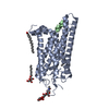
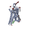
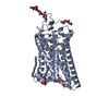
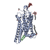
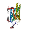
- PDB-4j4q: Crystal structure of active conformation of GPCR opsin stabilized... -
+ Open data

- Basic information
| Entry | Database: PDB / ID: 4j4q | |||||||||
|---|---|---|---|---|---|---|---|---|---|---|
| Title | Crystal structure of active conformation of GPCR opsin stabilized by octylglucoside | |||||||||
 Components Components |
| |||||||||
 Keywords Keywords |  SIGNALING PROTEIN / G-protein coupled receptor / Photoreceptor protein / Receptor Retinal protein / Transducer / Transmembrane / Glycoprotein / Lipoprotein / Sensory transduction / galpha subunit / GTP-binding / myristate / nucleotide-binding / Retinal Binding / Membrane SIGNALING PROTEIN / G-protein coupled receptor / Photoreceptor protein / Receptor Retinal protein / Transducer / Transmembrane / Glycoprotein / Lipoprotein / Sensory transduction / galpha subunit / GTP-binding / myristate / nucleotide-binding / Retinal Binding / Membrane | |||||||||
| Function / homology |  Function and homology informationOpsins / VxPx cargo-targeting to cilium / rod photoreceptor outer segment / rod bipolar cell differentiation / sperm head plasma membrane / negative regulation of cyclic-nucleotide phosphodiesterase activity / podosome assembly / absorption of visible light / opsin binding / The canonical retinoid cycle in rods (twilight vision) ...Opsins / VxPx cargo-targeting to cilium / rod photoreceptor outer segment / rod bipolar cell differentiation / sperm head plasma membrane / negative regulation of cyclic-nucleotide phosphodiesterase activity / podosome assembly / absorption of visible light / opsin binding / The canonical retinoid cycle in rods (twilight vision) / : / G protein-coupled photoreceptor activity / photoreceptor inner segment membrane / rhodopsin mediated signaling pathway / 11-cis retinal binding / cellular response to light stimulus / G protein-coupled receptor complex / Inactivation, recovery and regulation of the phototransduction cascade / phototransduction, visible light / thermotaxis / Activation of the phototransduction cascade / detection of temperature stimulus involved in thermoception / outer membrane / arrestin family protein binding / photoreceptor cell maintenance / acyl binding / photoreceptor outer segment membrane / G alpha (i) signalling events / response to light stimulus / phototransduction / photoreceptor outer segment / G-protein alpha-subunit binding / sperm midpiece / visual perception / photoreceptor inner segment / guanyl-nucleotide exchange factor activity / G protein-coupled receptor binding / G-protein beta/gamma-subunit complex binding / adenylate cyclase-modulating G protein-coupled receptor signaling pathway / microtubule cytoskeleton organization / photoreceptor disc membrane / GDP binding / heterotrimeric G-protein complex / cell-cell junction / gene expression / G protein-coupled receptor signaling pathway / Golgi membrane / GTPase activity / GTP binding / protein kinase binding / zinc ion binding / membrane / identical protein binding / metal ion binding / plasma membrane Function and homology informationOpsins / VxPx cargo-targeting to cilium / rod photoreceptor outer segment / rod bipolar cell differentiation / sperm head plasma membrane / negative regulation of cyclic-nucleotide phosphodiesterase activity / podosome assembly / absorption of visible light / opsin binding / The canonical retinoid cycle in rods (twilight vision) ...Opsins / VxPx cargo-targeting to cilium / rod photoreceptor outer segment / rod bipolar cell differentiation / sperm head plasma membrane / negative regulation of cyclic-nucleotide phosphodiesterase activity / podosome assembly / absorption of visible light / opsin binding / The canonical retinoid cycle in rods (twilight vision) / : / G protein-coupled photoreceptor activity / photoreceptor inner segment membrane / rhodopsin mediated signaling pathway / 11-cis retinal binding / cellular response to light stimulus / G protein-coupled receptor complex / Inactivation, recovery and regulation of the phototransduction cascade / phototransduction, visible light / thermotaxis / Activation of the phototransduction cascade / detection of temperature stimulus involved in thermoception / outer membrane / arrestin family protein binding / photoreceptor cell maintenance / acyl binding / photoreceptor outer segment membrane / G alpha (i) signalling events / response to light stimulus / phototransduction / photoreceptor outer segment / G-protein alpha-subunit binding / sperm midpiece / visual perception / photoreceptor inner segment / guanyl-nucleotide exchange factor activity / G protein-coupled receptor binding / G-protein beta/gamma-subunit complex binding / adenylate cyclase-modulating G protein-coupled receptor signaling pathway / microtubule cytoskeleton organization / photoreceptor disc membrane / GDP binding / heterotrimeric G-protein complex / cell-cell junction / gene expression / G protein-coupled receptor signaling pathway / Golgi membrane / GTPase activity / GTP binding / protein kinase binding / zinc ion binding / membrane / identical protein binding / metal ion binding / plasma membraneSimilarity search - Function | |||||||||
| Biological species |  Bos taurus (cattle) Bos taurus (cattle) | |||||||||
| Method | X-RAY DIFFRACTION / SYNCHROTRON / MOLECULAR REPLACEMENT / Resolution: 2.65 Å | |||||||||
 Authors Authors | Park, J.H. / Morizumi, T. / Li, Y. / Hong, J.E. / Pai, E.F. / Hofmann, K.P. / Choe, H.W. / Ernst, O.P. | |||||||||
 Citation Citation | Journal: Angew.Chem.Int.Ed.Engl. / Year: 2013 Title: Opsin, a structural model for olfactory receptors? Authors: Park, J.H. / Morizumi, T. / Li, Y. / Hong, J.E. / Pai, E.F. / Hofmann, K.P. / Choe, H.W. / Ernst, O.P. | |||||||||
| History |
|
- Structure visualization
| Structure viewer | Molecule: MolmilJmol/JSmol |
|---|
- Downloads & links
-Download
| PDBx/mmCIF format | 4j4q.cif.gz | 88.9 KB | Display | PDBx/mmCIF format |
|---|---|---|---|---|
| PDB format | pdb4j4q.ent.gz | 65.2 KB | Display | PDB format |
| PDBx/mmJSON format | 4j4q.json.gz | Tree view | PDBx/mmJSON format | |
| Others |  Other downloads Other downloads |
-Validation report
| Arichive directory | https://data.pdbj.org/pub/pdb/validation_reports/j4/4j4qftp://data.pdbj.org/pub/pdb/validation_reports/j4/4j4q | HTTPS FTP |
|---|
-Related structure data
| Related structure data | 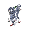 3dqbS S: Starting model for refinement |
|---|---|
| Similar structure data |


-Links
 PDBj
PDBj


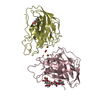
- Assembly
| Deposited unit | 
| ||||||||
|---|---|---|---|---|---|---|---|---|---|
| 1 |
| ||||||||
| Unit cell |
|
-Components
-Protein / Protein/peptide , 2 types, 2 molecules AB
| #1: Protein | Mass: 39031.457 Da / Num. of mol.: 1 / Source method: isolated from a natural source / Source: (natural) Bos taurus (cattle) / Strain: BovineBovinae / References: UniProt: P02699 |
|---|---|
| #2: Protein/peptide | Mass: 1261.487 Da / Num. of mol.: 1 / Fragment: C-terminal derived peptide, UNP RESIDUES 340-350 / Mutation: K341L, C347V / Source method: obtained synthetically Details: THIS PEPTIDE WAS CHEMICALLY SYNTHESIZED WITH K341L and C347V MUTATION Source: (synth.) Bos taurus (cattle) / References: UniProt: P04695 |
-Sugars , 3 types, 6 molecules 
| #3: Polysaccharide | alpha-D-mannopyranose-(1-3)-beta-D-mannopyranose-(1-4)-2-acetamido-2-deoxy-beta-D-glucopyranose-(1- ...alpha-D-mannopyranose-(1-3)-beta-D-mannopyranose-(1-4)-2-acetamido-2-deoxy-beta-D-glucopyranose-(1-4)-2-acetamido-2-deoxy-beta-D-glucopyranose / Mass: 748.682 Da / Num. of mol.: 1 Source method: isolated from a genetically manipulated source |
|---|---|
| #4: Polysaccharide | alpha-D-glucopyranose-(1-1)-alpha-D-glucopyranose / trehalose / 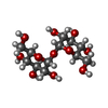  , Oligosaccharide / Class: Nutrient / Mass: 342.297 Da / Num. of mol.: 1 , Oligosaccharide / Class: Nutrient / Mass: 342.297 Da / Num. of mol.: 1Source method: isolated from a genetically manipulated source Details: oligosaccharide with reducing-end-to-reducing-end glycosidic bond References: trehalose |
| #5: Sugar | ChemComp-BOG / Octyl glucoside Type: D-saccharide / Mass: 292.369 Da / Num. of mol.: 4 Type: D-saccharide / Mass: 292.369 Da / Num. of mol.: 4Source method: isolated from a genetically manipulated source Formula: C14H28O6 / Comment: detergent*YM |
-Non-polymers , 4 types, 36 molecules 


| #6: Chemical | ChemComp-PLM / Palmitic acid Mass: 256.424 Da / Num. of mol.: 1 / Source method: obtained synthetically / Formula: C16H32O2 Mass: 256.424 Da / Num. of mol.: 1 / Source method: obtained synthetically / Formula: C16H32O2 | ||||
|---|---|---|---|---|---|
| #7: Chemical | Sulfate Mass: 96.063 Da / Num. of mol.: 2 / Source method: obtained synthetically / Formula: SO4 Mass: 96.063 Da / Num. of mol.: 2 / Source method: obtained synthetically / Formula: SO4#8: Chemical | ChemComp-ACT / | Acetate Mass: 59.044 Da / Num. of mol.: 1 / Source method: obtained synthetically / Formula: C2H3O2 Mass: 59.044 Da / Num. of mol.: 1 / Source method: obtained synthetically / Formula: C2H3O2#9: Water | ChemComp-HOH / | WaterMass: 18.015 Da / Num. of mol.: 32 / Source method: isolated from a natural source / Formula: H2O |
-Experimental details
-Experiment
| Experiment | Method: X-RAY DIFFRACTION / Number of used crystals: 1 |
|---|
- Sample preparation
| Crystal grow | Temperature: 277 K / Method: vapor diffusion, hanging drop / pH: 5.6 Details: AMMONIUM SULFATE, pH 5.6, VAPOR DIFFUSION, HANGING DROP, temperature 277K |
|---|
-Data collection
| Diffraction | Mean temperature: 100 K |
|---|---|
| Diffraction source | Source: SYNCHROTRON / Site: BESSY  / Beamline: 14.2 / Wavelength: 0.91841 Å / Beamline: 14.2 / Wavelength: 0.91841 Å |
| Detector | Type: MARMOSAIC 225 mm CCD / Detector: CCD / Date: May 6, 2010 / Details: MIRRORS |
| Radiation | Monochromator: SI-111 CRYSTAL-DOUBLE CRYSTAL MONOCHROMATOR / Protocol: SINGLE WAVELENGTH / Monochromatic (M) / Laue (L): M / Scattering type: x-ray |
| Radiation wavelength | Wavelength: 0.91841 Å / Relative weight: 1 |
| Reflection | Resolution: 2.65→33.46 Å / Num. all: 35401 / Num. obs: 35401 / % possible obs: 99.95 % / Observed criterion σ(F): 0 / Observed criterion σ(I): 0 / Redundancy: 4.4 % / Rmerge(I) obs: 0.055 / Rsym value: 0.055 / Net I/σ(I): 17.3 |
| Reflection shell | Resolution: 2.65→2.79 Å / Redundancy: 4.5 % / Rmerge(I) obs: 0.421 / Mean I/σ(I) obs: 3.3 / Rsym value: 0.421 / % possible all: 100 |
- Processing
| Software |
| ||||||||||||||||||||||||||||||||||||||||||||||||||||||||||||||||||||||||||||||||||||||||||||||||||||||
|---|---|---|---|---|---|---|---|---|---|---|---|---|---|---|---|---|---|---|---|---|---|---|---|---|---|---|---|---|---|---|---|---|---|---|---|---|---|---|---|---|---|---|---|---|---|---|---|---|---|---|---|---|---|---|---|---|---|---|---|---|---|---|---|---|---|---|---|---|---|---|---|---|---|---|---|---|---|---|---|---|---|---|---|---|---|---|---|---|---|---|---|---|---|---|---|---|---|---|---|---|---|---|---|
| Refinement | Method to determine structure: MOLECULAR REPLACEMENT Starting model: PDB ENTRY 3DQB Resolution: 2.65→33.46 Å / Isotropic thermal model: Anisotropic / Cross valid method: THROUGHOUT / Stereochemistry target values: MAXIMUM LIKELIHOOD / Details: HYDROGENS HAVE BEEN ADDED IN THE RIDING POSITIONS
| ||||||||||||||||||||||||||||||||||||||||||||||||||||||||||||||||||||||||||||||||||||||||||||||||||||||
| Solvent computation | Ion probe radii: 0.8 Å / Shrinkage radii: 0.8 Å / VDW probe radii: 1.2 Å | ||||||||||||||||||||||||||||||||||||||||||||||||||||||||||||||||||||||||||||||||||||||||||||||||||||||
| Displacement parameters | Biso mean: 55.824 Å2
| ||||||||||||||||||||||||||||||||||||||||||||||||||||||||||||||||||||||||||||||||||||||||||||||||||||||
| Refinement step | Cycle: LAST / Resolution: 2.65→33.46 Å
| ||||||||||||||||||||||||||||||||||||||||||||||||||||||||||||||||||||||||||||||||||||||||||||||||||||||
| Refine LS restraints |
| ||||||||||||||||||||||||||||||||||||||||||||||||||||||||||||||||||||||||||||||||||||||||||||||||||||||
| LS refinement shell | Resolution: 2.65→2.718 Å / Total num. of bins used: 20
|
