 Movie
Movie Controller

[English] 日本語
 Yorodumi
- PDB-4j1b: X-ray structure of the adduct between hen egg white lysozyme and ... -
+ Open data

- Basic information
| Entry | Database: PDB / ID: 4j1b | ||||||
|---|---|---|---|---|---|---|---|
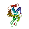
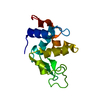
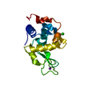
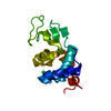
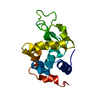
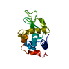
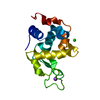
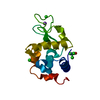
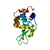
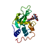
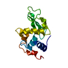
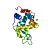
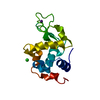
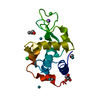
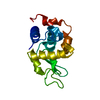
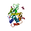
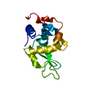
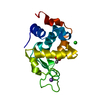
| Title | X-ray structure of the adduct between hen egg white lysozyme and AziRu (black crystal) | ||||||
 Components Components | Lysozyme C | ||||||
 Keywords Keywords |  HYDROLASE / C-type lysozyme/alpha-lactalbumin family HYDROLASE / C-type lysozyme/alpha-lactalbumin family | ||||||
| Function / homology |  Function and homology informationAntimicrobial peptides / Neutrophil degranulation / beta-N-acetylglucosaminidase activity / cell wall macromolecule catabolic process / lysozyme / lysozyme activity / killing of cells of another organism / defense response to Gram-negative bacterium / defense response to Gram-positive bacterium / defense response to bacterium ...Antimicrobial peptides / Neutrophil degranulation / beta-N-acetylglucosaminidase activity / cell wall macromolecule catabolic process / lysozyme / lysozyme activity / killing of cells of another organism / defense response to Gram-negative bacterium / defense response to Gram-positive bacterium / defense response to bacterium / endoplasmic reticulum / extracellular space / identical protein binding / cytoplasm Function and homology informationAntimicrobial peptides / Neutrophil degranulation / beta-N-acetylglucosaminidase activity / cell wall macromolecule catabolic process / lysozyme / lysozyme activity / killing of cells of another organism / defense response to Gram-negative bacterium / defense response to Gram-positive bacterium / defense response to bacterium ...Antimicrobial peptides / Neutrophil degranulation / beta-N-acetylglucosaminidase activity / cell wall macromolecule catabolic process / lysozyme / lysozyme activity / killing of cells of another organism / defense response to Gram-negative bacterium / defense response to Gram-positive bacterium / defense response to bacterium / endoplasmic reticulum / extracellular space / identical protein binding / cytoplasmSimilarity search - Function | ||||||
| Biological species |  Gallus gallus (chicken) Gallus gallus (chicken) | ||||||
| Method | X-RAY DIFFRACTION / FOURIER SYNTHESIS / Resolution: 1.66 Å | ||||||
 Authors Authors | Vergara, A. / Merlino, A. | ||||||
 Citation Citation | Journal: Inorg.Chem. / Year: 2013 Title: Interaction of Anticancer Ruthenium Compounds with Proteins: High-Resolution X-ray Structures and Raman Microscopy Studies of the Adduct between Hen Egg White Lysozyme and AziRu. Authors: Vergara, A. / D'Errico, G. / Montesarchio, D. / Mangiapia, G. / Paduano, L. / Merlino, A. | ||||||
| History |
|
- Structure visualization
| Structure viewer | Molecule: MolmilJmol/JSmol |
|---|
- Downloads & links
-Download
| PDBx/mmCIF format | 4j1b.cif.gz | 39.8 KB | Display | PDBx/mmCIF format |
|---|---|---|---|---|
| PDB format | pdb4j1b.ent.gz | 30.4 KB | Display | PDB format |
| PDBx/mmJSON format | 4j1b.json.gz | Tree view | PDBx/mmJSON format | |
| Others |  Other downloads Other downloads |
-Validation report
| Arichive directory | https://data.pdbj.org/pub/pdb/validation_reports/j1/4j1bftp://data.pdbj.org/pub/pdb/validation_reports/j1/4j1b | HTTPS FTP |
|---|
-Related structure data


-Links
 PDBj
PDBj


- Assembly
| Deposited unit | 
| |||||||||
|---|---|---|---|---|---|---|---|---|---|---|
| 1 |
| |||||||||
| Unit cell |
| |||||||||
| Components on special symmetry positions |
|
-Components
| #1: Protein | Mass: 14331.160 Da / Num. of mol.: 1 / Source method: isolated from a natural source / Source: (natural) Gallus gallus (chicken) / References: UniProt: P00698, lysozyme | ||||||||
|---|---|---|---|---|---|---|---|---|---|
| #2: Chemical | Chloride  Mass: 35.453 Da / Num. of mol.: 2 / Source method: obtained synthetically / Formula: Cl Mass: 35.453 Da / Num. of mol.: 2 / Source method: obtained synthetically / Formula: Cl#3: Chemical | ChemComp-NA / |   Mass: 22.990 Da / Num. of mol.: 1 / Source method: obtained synthetically / Formula: Na Mass: 22.990 Da / Num. of mol.: 1 / Source method: obtained synthetically / Formula: Na#4: Chemical | ChemComp-RU / | Ruthenium  Mass: 101.070 Da / Num. of mol.: 1 / Source method: obtained synthetically / Formula: Ru Mass: 101.070 Da / Num. of mol.: 1 / Source method: obtained synthetically / Formula: Ru#5: Water | ChemComp-HOH / | Water Mass: 18.015 Da / Num. of mol.: 129 / Source method: isolated from a natural source / Formula: H2O Mass: 18.015 Da / Num. of mol.: 129 / Source method: isolated from a natural source / Formula: H2ONonpolymer details | HEN EGG WHITE LYSOZYME WAS CRYSTALLIZED WITH AN ORGANO-METALLIC RUTHENIUM(III) COMPOUND, CALLED ...HEN EGG WHITE LYSOZYME WAS CRYSTALLIZ | |
-Experimental details
-Experiment
| Experiment | Method: X-RAY DIFFRACTION / Number of used crystals: 1 |
|---|
- Sample preparation
| Crystal | Density Matthews: 1.97 Å3/Da / Density % sol: 37.69 % |
|---|---|
| Crystal grow | Temperature: 293 K / Method: vapor diffusion, hanging drop / pH: 4.5 Details: 1.0-1.4 M NaCl, 0.050 M acetate buffer.Crystals were soaked in a saturated solution of AziRu containing 2.4 M NaCl in the same buffer. After a few hours, crystals changed their colour to ...Details: 1.0-1.4 M NaCl, 0.050 M acetate buffer.Crystals were soaked in a saturated solution of AziRu containing 2.4 M NaCl in the same buffer. After a few hours, crystals changed their colour to yellow and then green. In a few days they turned to black, pH 4.5, VAPOR DIFFUSION, HANGING DROP, temperature 293K |
-Data collection
| Diffraction | Mean temperature: 100 K |
|---|---|
| Diffraction source | Source: ROTATING ANODE / Type: RIGAKU MICROMAX-007 HF / Wavelength: 1.5418 Å |
| Detector | Type: RIGAKU SATURN 944 / Detector: CCD |
| Radiation | Monochromator: graphite / Protocol: SINGLE WAVELENGTH / Monochromatic (M) / Laue (L): M / Scattering type: x-ray |
| Radiation wavelength | Wavelength: 1.5418 Å / Relative weight: 1 |
| Reflection | Resolution: 1.66→50 Å / Num. obs: 13847 / % possible obs: 97.9 % / Observed criterion σ(F): 0 / Observed criterion σ(I): 0 / Biso Wilson estimate: 23.3 Å2 |
- Processing
| Software |
| ||||||||||||||||||||||||||||||||||||||||||||||||||||||||||||||||||||||||||||||||
|---|---|---|---|---|---|---|---|---|---|---|---|---|---|---|---|---|---|---|---|---|---|---|---|---|---|---|---|---|---|---|---|---|---|---|---|---|---|---|---|---|---|---|---|---|---|---|---|---|---|---|---|---|---|---|---|---|---|---|---|---|---|---|---|---|---|---|---|---|---|---|---|---|---|---|---|---|---|---|---|---|---|
| Refinement | Method to determine structure: FOURIER SYNTHESIS / Resolution: 1.66→33.57 Å / Rfactor Rfree error: 0.009 / Data cutoff high absF: 300954.92 / Data cutoff low absF: 0 / Isotropic thermal model: RESTRAINED / Cross valid method: THROUGHOUT / σ(F): 2 / Details: BULK SOLVENT MODEL USED
| ||||||||||||||||||||||||||||||||||||||||||||||||||||||||||||||||||||||||||||||||
| Solvent computation | Solvent model: FLAT MODEL / Bsol: 56.1439 Å2 / ksol: 0.38 e/Å3 | ||||||||||||||||||||||||||||||||||||||||||||||||||||||||||||||||||||||||||||||||
| Displacement parameters | Biso mean: 25.3 Å2
| ||||||||||||||||||||||||||||||||||||||||||||||||||||||||||||||||||||||||||||||||
| Refine analyze |
| ||||||||||||||||||||||||||||||||||||||||||||||||||||||||||||||||||||||||||||||||
| Refinement step | Cycle: LAST / Resolution: 1.66→33.57 Å
| ||||||||||||||||||||||||||||||||||||||||||||||||||||||||||||||||||||||||||||||||
| Refine LS restraints |
| ||||||||||||||||||||||||||||||||||||||||||||||||||||||||||||||||||||||||||||||||
| Refine LS restraints NCS | NCS model details: NONE | ||||||||||||||||||||||||||||||||||||||||||||||||||||||||||||||||||||||||||||||||
| LS refinement shell | Resolution: 1.66→1.76 Å / Rfactor Rfree error: 0.028 / Total num. of bins used: 6
|
