 Movie
Movie Controller


+ Open data

- Basic information
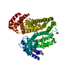
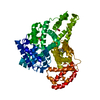
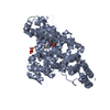
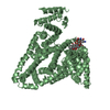
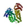
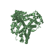
| Entry | Database: PDB / ID: 4iw2 | ||||||
|---|---|---|---|---|---|---|---|
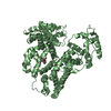
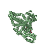
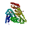
| Title | HSA-glucose complex | ||||||
 Components Components | Serum albumin | ||||||
 Keywords Keywords | TRANSPORT PROTEIN / Heart shape / tansport | ||||||
| Function / homology |  Function and homology information Function and homology informationcellular response to calcium ion starvation / exogenous protein binding / Ciprofloxacin ADME / HDL remodeling / enterobactin binding / Heme biosynthesis / negative regulation of mitochondrial depolarization / Prednisone ADME / Heme degradation / antioxidant activity ...cellular response to calcium ion starvation / exogenous protein binding / Ciprofloxacin ADME / HDL remodeling / enterobactin binding / Heme biosynthesis / negative regulation of mitochondrial depolarization / Prednisone ADME / Heme degradation / antioxidant activity / Aspirin ADME / toxic substance binding / Scavenging of heme from plasma / Recycling of bile acids and salts / cellular response to starvation / platelet alpha granule lumen / fatty acid binding / Post-translational protein phosphorylation / Cytoprotection by HMOX1 / Regulation of Insulin-like Growth Factor (IGF) transport and uptake by Insulin-like Growth Factor Binding Proteins (IGFBPs) / pyridoxal phosphate binding / Platelet degranulation / protein-folding chaperone binding / blood microparticle / copper ion binding / endoplasmic reticulum lumen / Golgi apparatus / endoplasmic reticulum / protein-containing complex / DNA binding / extracellular space / extracellular exosome / extracellular region / identical protein binding / nucleus / cytoplasmSimilarity search - Function | ||||||
| Biological species |  Homo sapiens (human) Homo sapiens (human) | ||||||
| Method | X-RAY DIFFRACTION / SYNCHROTRON / MOLECULAR REPLACEMENT / Resolution: 2.41 Å | ||||||
 Authors Authors | Wang, Y. / Yu, H. / Shi, X. / Luo, Z. / Huang, M. | ||||||
 Citation Citation | Journal: J.Biol.Chem. / Year: 2013 Title: Structural mechanism of ring-opening reaction of glucose by human serum albumin Authors: Wang, Y. / Yu, H. / Shi, X. / Luo, Z. / Lin, D. / Huang, M. | ||||||
| History |
|
- Structure visualization
| Structure viewer | Molecule: MolmilJmol/JSmol |
|---|
- Downloads & links
-Download
| PDBx/mmCIF format | 4iw2.cif.gz | 126 KB | Display | PDBx/mmCIF format |
|---|---|---|---|---|
| PDB format | pdb4iw2.ent.gz | 97.1 KB | Display | PDB format |
| PDBx/mmJSON format | 4iw2.json.gz | Tree view | PDBx/mmJSON format | |
| Others |  Other downloads Other downloads |
-Validation report
| Arichive directory | https://data.pdbj.org/pub/pdb/validation_reports/iw/4iw2ftp://data.pdbj.org/pub/pdb/validation_reports/iw/4iw2 | HTTPS FTP |
|---|
-Related structure data
| Related structure data | 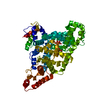 4iw1C  4k2cC  1ao6S C: citing same article ( S: Starting model for refinement |
|---|---|
| Similar structure data |


-Links
 PDBj
PDBj


- Assembly
| Deposited unit | 
| ||||||||
|---|---|---|---|---|---|---|---|---|---|
| 1 |
| ||||||||
| Unit cell |
|
-Components
| #1: Protein | Mass: 66571.219 Da / Num. of mol.: 1 Source method: isolated from a genetically manipulated source Source: (gene. exp.) Homo sapiens (human) / Gene: ALB / Production host:  Pichia pastoris (fungus) / References: UniProt: P02768 Pichia pastoris (fungus) / References: UniProt: P02768 |
|---|---|
| #2: Sugar | ChemComp-GLO / Glucose  Type: D-saccharide / Mass: 180.156 Da / Num. of mol.: 1 Type: D-saccharide / Mass: 180.156 Da / Num. of mol.: 1Source method: isolated from a genetically manipulated source Formula: C6H12O6 |
| #3: Sugar | ChemComp-GLC / Glucose  Type: D-saccharide, alpha linking / Mass: 180.156 Da / Num. of mol.: 1 Type: D-saccharide, alpha linking / Mass: 180.156 Da / Num. of mol.: 1Source method: isolated from a genetically manipulated source Formula: C6H12O6 |
| #4: Chemical | ChemComp-PO4 / Phosphate  Mass: 94.971 Da / Num. of mol.: 1 / Source method: obtained synthetically / Formula: PO4 Mass: 94.971 Da / Num. of mol.: 1 / Source method: obtained synthetically / Formula: PO4 |
| #5: Water | ChemComp-HOH / Water Mass: 18.015 Da / Num. of mol.: 6 / Source method: isolated from a natural source / Formula: H2O Mass: 18.015 Da / Num. of mol.: 6 / Source method: isolated from a natural source / Formula: H2O |
-Experimental details
-Experiment
| Experiment | Method: X-RAY DIFFRACTION / Number of used crystals: 1 |
|---|
- Sample preparation
| Crystal | Density Matthews: 2.13 Å3/Da / Density % sol: 42.38 % |
|---|---|
| Crystal grow | Temperature: 298 K / Method: vapor diffusion, sitting drop / pH: 7.4 Details: 25%-30% PEG 3350, 50mM potassium phosphate, pH 7.4, VAPOR DIFFUSION, SITTING DROP, temperature 298K |
-Data collection
| Diffraction | Mean temperature: 298 K |
|---|---|
| Diffraction source | Source: SYNCHROTRON / Site: APS  / Beamline: 17-ID / Beamline: 17-ID |
| Detector | Type: ADSC QUANTUM 210 / Detector: CCD / Date: Oct 1, 2012 |
| Radiation | Protocol: SINGLE WAVELENGTH / Monochromatic (M) / Laue (L): M / Scattering type: x-ray |
| Radiation wavelength | Relative weight: 1 |
| Reflection | Resolution: 2.4→50 Å / Num. obs: 19250 / Biso Wilson estimate: 27.8 Å2 |
- Processing
| Software |
| ||||||||||||||||||||||||||||||||||||||||||||||||||||||||||||
|---|---|---|---|---|---|---|---|---|---|---|---|---|---|---|---|---|---|---|---|---|---|---|---|---|---|---|---|---|---|---|---|---|---|---|---|---|---|---|---|---|---|---|---|---|---|---|---|---|---|---|---|---|---|---|---|---|---|---|---|---|---|
| Refinement | Method to determine structure: MOLECULAR REPLACEMENT Starting model: 1AO6 Resolution: 2.41→33.4 Å / Rfactor Rfree error: 0.006 / Data cutoff high absF: 103937.05 / Data cutoff low absF: 0 / Isotropic thermal model: RESTRAINED / Cross valid method: THROUGHOUT / σ(F): 0 / Details: BULK SOLVENT MODEL USED
| ||||||||||||||||||||||||||||||||||||||||||||||||||||||||||||
| Solvent computation | Solvent model: FLAT MODEL / Bsol: 36.4208 Å2 / ksol: 0.32 e/Å3 | ||||||||||||||||||||||||||||||||||||||||||||||||||||||||||||
| Displacement parameters | Biso mean: 58.2 Å2
| ||||||||||||||||||||||||||||||||||||||||||||||||||||||||||||
| Refine analyze |
| ||||||||||||||||||||||||||||||||||||||||||||||||||||||||||||
| Refinement step | Cycle: LAST / Resolution: 2.41→33.4 Å
| ||||||||||||||||||||||||||||||||||||||||||||||||||||||||||||
| Refine LS restraints |
| ||||||||||||||||||||||||||||||||||||||||||||||||||||||||||||
| LS refinement shell | Resolution: 2.4→2.55 Å / Rfactor Rfree error: 0.021 / Total num. of bins used: 6
| ||||||||||||||||||||||||||||||||||||||||||||||||||||||||||||
| Xplor file |
|
