 Movie
Movie Controller

[English] 日本語
 Yorodumi
- PDB-4ipx: Analyzing the visible conformational substates of the FK506 bindi... -
+ Open data

- Basic information
| Entry | Database: PDB / ID: 4ipx | ||||||
|---|---|---|---|---|---|---|---|
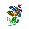
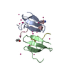
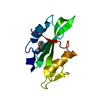
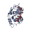
| Title | Analyzing the visible conformational substates of the FK506 binding protein FKBP12 | ||||||
 Components Components | Peptidyl-prolyl cis-trans isomerase FKBP1A | ||||||
 Keywords Keywords |  ISOMERASE ISOMERASE | ||||||
| Function / homology |  Function and homology informationmacrolide binding / activin receptor binding / cytoplasmic side of membrane / transforming growth factor beta receptor binding / TGFBR1 LBD Mutants in Cancer / signaling receptor inhibitor activity / type I transforming growth factor beta receptor binding / negative regulation of activin receptor signaling pathway / regulation of amyloid precursor protein catabolic process / heart trabecula formation ...macrolide binding / activin receptor binding / cytoplasmic side of membrane / transforming growth factor beta receptor binding / TGFBR1 LBD Mutants in Cancer / signaling receptor inhibitor activity / type I transforming growth factor beta receptor binding / negative regulation of activin receptor signaling pathway / regulation of amyloid precursor protein catabolic process / heart trabecula formation / terminal cisterna / ryanodine receptor complex / I-SMAD binding / protein maturation by protein folding / 'de novo' protein folding / ventricular cardiac muscle tissue morphogenesis / negative regulation of phosphoprotein phosphatase activity / FK506 binding / mTORC1-mediated signalling / TGF-beta receptor signaling activates SMADs / Calcineurin activates NFAT / regulation of immune response / protein peptidyl-prolyl isomerization / supramolecular fiber organization / heart morphogenesis / regulation of ryanodine-sensitive calcium-release channel activity / sarcoplasmic reticulum membrane / T cell activation / TGF-beta receptor signaling in EMT (epithelial to mesenchymal transition) / sarcoplasmic reticulum / peptidylprolyl isomerase / peptidyl-prolyl cis-trans isomerase activity / calcium ion transmembrane transport / negative regulation of transforming growth factor beta receptor signaling pathway / Z disc / SARS-CoV-1 activates/modulates innate immune responses / regulation of protein localization / protein folding / positive regulation of protein binding / protein refolding / positive regulation of canonical NF-kappaB signal transduction / amyloid fibril formation / Potential therapeutics for SARS / transmembrane transporter binding / membrane / cytosol / cytoplasm Function and homology informationmacrolide binding / activin receptor binding / cytoplasmic side of membrane / transforming growth factor beta receptor binding / TGFBR1 LBD Mutants in Cancer / signaling receptor inhibitor activity / type I transforming growth factor beta receptor binding / negative regulation of activin receptor signaling pathway / regulation of amyloid precursor protein catabolic process / heart trabecula formation ...macrolide binding / activin receptor binding / cytoplasmic side of membrane / transforming growth factor beta receptor binding / TGFBR1 LBD Mutants in Cancer / signaling receptor inhibitor activity / type I transforming growth factor beta receptor binding / negative regulation of activin receptor signaling pathway / regulation of amyloid precursor protein catabolic process / heart trabecula formation / terminal cisterna / ryanodine receptor complex / I-SMAD binding / protein maturation by protein folding / 'de novo' protein folding / ventricular cardiac muscle tissue morphogenesis / negative regulation of phosphoprotein phosphatase activity / FK506 binding / mTORC1-mediated signalling / TGF-beta receptor signaling activates SMADs / Calcineurin activates NFAT / regulation of immune response / protein peptidyl-prolyl isomerization / supramolecular fiber organization / heart morphogenesis / regulation of ryanodine-sensitive calcium-release channel activity / sarcoplasmic reticulum membrane / T cell activation / TGF-beta receptor signaling in EMT (epithelial to mesenchymal transition) / sarcoplasmic reticulum / peptidylprolyl isomerase / peptidyl-prolyl cis-trans isomerase activity / calcium ion transmembrane transport / negative regulation of transforming growth factor beta receptor signaling pathway / Z disc / SARS-CoV-1 activates/modulates innate immune responses / regulation of protein localization / protein folding / positive regulation of protein binding / protein refolding / positive regulation of canonical NF-kappaB signal transduction / amyloid fibril formation / Potential therapeutics for SARS / transmembrane transporter binding / membrane / cytosol / cytoplasmSimilarity search - Function | ||||||
| Biological species |  Homo sapiens (human) Homo sapiens (human) | ||||||
| Method | X-RAY DIFFRACTION / MOLECULAR REPLACEMENT / Resolution: 1.7 Å | ||||||
 Authors Authors | Chen, H. / Mustafi, S.M. / Li, H.M. / LeMaster, D.M. / Hernandez, G. | ||||||
 Citation Citation | Journal: Biochem.J. / Year: 2013 Title: Analysing the visible conformational substates of the FK506-binding protein FKBP12. Authors: Mustafi, S.M. / Chen, H. / Li, H. / Lemaster, D.M. / Hernandez, G. | ||||||
| History |
|
- Structure visualization
| Structure viewer | Molecule: MolmilJmol/JSmol |
|---|
- Downloads & links
-Download
| PDBx/mmCIF format | 4ipx.cif.gz | 36.5 KB | Display | PDBx/mmCIF format |
|---|---|---|---|---|
| PDB format | pdb4ipx.ent.gz | 24 KB | Display | PDB format |
| PDBx/mmJSON format | 4ipx.json.gz | Tree view | PDBx/mmJSON format | |
| Others |  Other downloads Other downloads |
-Validation report
| Arichive directory | https://data.pdbj.org/pub/pdb/validation_reports/ip/4ipxftp://data.pdbj.org/pub/pdb/validation_reports/ip/4ipx | HTTPS FTP |
|---|
-Related structure data
| Related structure data |  2ppnS S: Starting model for refinement |
|---|---|
| Similar structure data |


-Links
 PDBj
PDBj


- Assembly
| Deposited unit | 
| |||||||||||||||
|---|---|---|---|---|---|---|---|---|---|---|---|---|---|---|---|---|
| 1 |
| |||||||||||||||
| Unit cell |
| |||||||||||||||
| Components on special symmetry positions |
|
-Components
| #1: Protein | Mass: 11793.480 Da / Num. of mol.: 1 / Mutation: C22V, H87V Source method: isolated from a genetically manipulated source Source: (gene. exp.) Homo sapiens (human) / Gene: FKBP1A, FKBP1, FKBP12 / Production host:  Escherichia coli (E. coli) / References: UniProt: P62942, peptidylprolyl isomerase Escherichia coli (E. coli) / References: UniProt: P62942, peptidylprolyl isomerase | ||
|---|---|---|---|
| #2: Chemical | 2-Methyl-2,4-pentanediol  Mass: 118.174 Da / Num. of mol.: 2 / Source method: obtained synthetically / Formula: C6H14O2 / Comment: precipitant*YM Mass: 118.174 Da / Num. of mol.: 2 / Source method: obtained synthetically / Formula: C6H14O2 / Comment: precipitant*YM#3: Water | ChemComp-HOH / | Water Mass: 18.015 Da / Num. of mol.: 127 / Source method: isolated from a natural source / Formula: H2O Mass: 18.015 Da / Num. of mol.: 127 / Source method: isolated from a natural source / Formula: H2O |
-Experimental details
-Experiment
| Experiment | Method: X-RAY DIFFRACTION / Number of used crystals: 1 |
|---|
- Sample preparation
| Crystal | Density Matthews: 2.19 Å3/Da / Density % sol: 43.8 % |
|---|---|
| Crystal grow | Temperature: 298 K / Method: vapor diffusion, hanging drop / pH: 7.4 Details: 1.7 M sodium malonate, pH 7.0, 0.1 M HEPES, pH 7.4, 5% MPD, VAPOR DIFFUSION, HANGING DROP, temperature 298K |
-Data collection
| Diffraction | Mean temperature: 100 K |
|---|---|
| Diffraction source | Source: ROTATING ANODE / Type: Cu FINE FOCUS / Wavelength: 1.5418 Å |
| Detector | Type: RIGAKU RAXIS IV++ / Detector: IMAGE PLATE / Date: Oct 28, 2012 |
| Radiation | Monochromator: mirror / Protocol: SINGLE WAVELENGTH / Monochromatic (M) / Laue (L): M / Scattering type: x-ray |
| Radiation wavelength | Wavelength: 1.5418 Å / Relative weight: 1 |
| Reflection | Resolution: 1.7→35 Å / Num. obs: 10996 / % possible obs: 93.7 % / Observed criterion σ(F): 1 / Observed criterion σ(I): 1 / Redundancy: 3.23 % / Rmerge(I) obs: 0.097 / Rsym value: 0.097 / Net I/σ(I): 29.4 |
| Reflection shell | Resolution: 1.7→1.76 Å / Redundancy: 3.4 % / Rmerge(I) obs: 0.296 / Mean I/σ(I) obs: 6.3 / Rsym value: 0.296 / % possible all: 93.6 |
- Processing
| Software |
| |||||||||||||||||||||||||||||||||||
|---|---|---|---|---|---|---|---|---|---|---|---|---|---|---|---|---|---|---|---|---|---|---|---|---|---|---|---|---|---|---|---|---|---|---|---|---|
| Refinement | Method to determine structure: MOLECULAR REPLACEMENT Starting model: PDB ENTRY 2PPN Resolution: 1.7→19.607 Å / SU ML: 0.31 / σ(F): 1.55 / Phase error: 23.8 / Stereochemistry target values: ML
| |||||||||||||||||||||||||||||||||||
| Solvent computation | Shrinkage radii: 0.47 Å / VDW probe radii: 0.8 Å / Solvent model: FLAT BULK SOLVENT MODEL / Bsol: 56.491 Å2 / ksol: 0.421 e/Å3 | |||||||||||||||||||||||||||||||||||
| Displacement parameters |
| |||||||||||||||||||||||||||||||||||
| Refinement step | Cycle: LAST / Resolution: 1.7→19.607 Å
| |||||||||||||||||||||||||||||||||||
| Refine LS restraints |
| |||||||||||||||||||||||||||||||||||
| LS refinement shell |
|
