 Movie
Movie Controller


+ Open data

- Basic information
| Entry | Database: PDB / ID: 4ii7 | ||||||
|---|---|---|---|---|---|---|---|
| Title | Archaellum Assembly ATPase FlaI | ||||||
 Components Components | FlaI ATPase | ||||||
 Keywords Keywords |  HYDROLASE / Hexamer / ATP/ADP / cytoplasmic / membrane associated HYDROLASE / Hexamer / ATP/ADP / cytoplasmic / membrane associated | ||||||
| Function / homology |  Function and homology information Function and homology information | ||||||
| Biological species |   Sulfolobus acidocaldarius (acidophilic) Sulfolobus acidocaldarius (acidophilic) | ||||||
| Method | X-RAY DIFFRACTION / SYNCHROTRON / MOLECULAR REPLACEMENT / Resolution: 3.59 Å | ||||||
 Authors Authors | Reindl, S. / Williams, G.J. / Tainer, J.A. | ||||||
 Citation Citation | Journal: Mol.Cell / Year: 2013 Title: Insights into FlaI Functions in Archaeal Motor Assembly and Motility from Structures, Conformations, and Genetics. Authors: Reindl, S. / Ghosh, A. / Williams, G.J. / Lassak, K. / Neiner, T. / Henche, A.L. / Albers, S.V. / Tainer, J.A. | ||||||
| History |
|
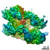
- Structure visualization
| Structure viewer | Molecule: MolmilJmol/JSmol |
|---|
- Downloads & links
-Download
| PDBx/mmCIF format | 4ii7.cif.gz | 395.4 KB | Display | PDBx/mmCIF format |
|---|---|---|---|---|
| PDB format | pdb4ii7.ent.gz | 329.3 KB | Display | PDB format |
| PDBx/mmJSON format | 4ii7.json.gz | Tree view | PDBx/mmJSON format | |
| Others |  Other downloads Other downloads |
-Validation report
| Arichive directory | https://data.pdbj.org/pub/pdb/validation_reports/ii/4ii7ftp://data.pdbj.org/pub/pdb/validation_reports/ii/4ii7 | HTTPS FTP |
|---|
-Related structure data
| Related structure data |  4ihqSC S: Starting model for refinement C: citing same article ( |
|---|---|
| Similar structure data |


-Links
 PDBj
PDBj
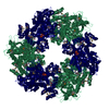
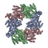
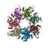
- Assembly
| Deposited unit | 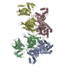
| ||||||||
|---|---|---|---|---|---|---|---|---|---|
| 1 | 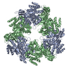
| ||||||||
| 2 | 
| ||||||||
| Unit cell |
|
-Components
| #1: Protein | Mass: 58835.430 Da / Num. of mol.: 4 Source method: isolated from a genetically manipulated source Source: (gene. exp.) Sulfolobus acidocaldarius (acidophilic)Strain: ATCC 33909 / DSM 639 / JCM 8929 / NBRC 15157 / NCIMB 11770 Gene: 1173, Saci_1173 / Production host:  Escherichia coli (E. coli) / References: UniProt: Q4J9L0, EC: 3.6.1.4 Escherichia coli (E. coli) / References: UniProt: Q4J9L0, EC: 3.6.1.4 |
|---|
-Experimental details
-Experiment
| Experiment | Method: X-RAY DIFFRACTION / Number of used crystals: 1 |
|---|
- Sample preparation
| Crystal | Density Matthews: 3.23 Å3/Da / Density % sol: 61.95 % |
|---|---|
| Crystal grow | Temperature: 293 K / pH: 7 Details: 100mM HEPES pH 7.0, 22% Polyvinylpyrrolidone K 15, 10 mM CoCl2, 15% glycerol , VAPOR DIFFUSION, HANGING DROP, temperature 293K |
-Data collection
| Diffraction | Mean temperature: 100 K |
|---|---|
| Diffraction source | Source: SYNCHROTRON / Site: ALS  / Beamline: 12.3.1 / Wavelength: 1.07 / Beamline: 12.3.1 / Wavelength: 1.07 |
| Detector | Type: ADSC QUANTUM 315r / Detector: CCD / Date: Oct 25, 2009 |
| Radiation | Monochromator: SI (111) / Protocol: SINGLE WAVELENGTH / Monochromatic (M) / Laue (L): M / Scattering type: x-ray |
| Radiation wavelength | Wavelength: 1.07 Å / Relative weight: 1 |
| Reflection | Resolution: 3.59→49.923 Å / Num. obs: 34740 / % possible obs: 99 % / Observed criterion σ(I): 1 |
| Reflection shell | Resolution: 3.59→3.76 Å / % possible all: 87.7 |
- Processing
| Software |
| |||||||||||||||||||||||||||||||||||||||||||||||||||||||||||||||||||||||||||||||||||||||||||||||||||||||||
|---|---|---|---|---|---|---|---|---|---|---|---|---|---|---|---|---|---|---|---|---|---|---|---|---|---|---|---|---|---|---|---|---|---|---|---|---|---|---|---|---|---|---|---|---|---|---|---|---|---|---|---|---|---|---|---|---|---|---|---|---|---|---|---|---|---|---|---|---|---|---|---|---|---|---|---|---|---|---|---|---|---|---|---|---|---|---|---|---|---|---|---|---|---|---|---|---|---|---|---|---|---|---|---|---|---|---|
| Refinement | Method to determine structure: MOLECULAR REPLACEMENT Starting model: 4IHQ Resolution: 3.59→49.92 Å / SU ML: 0.59 / σ(F): 1.35 / Phase error: 35.94 / Stereochemistry target values: ML
| |||||||||||||||||||||||||||||||||||||||||||||||||||||||||||||||||||||||||||||||||||||||||||||||||||||||||
| Solvent computation | Shrinkage radii: 0.9 Å / VDW probe radii: 1.11 Å / Solvent model: FLAT BULK SOLVENT MODEL | |||||||||||||||||||||||||||||||||||||||||||||||||||||||||||||||||||||||||||||||||||||||||||||||||||||||||
| Refinement step | Cycle: LAST / Resolution: 3.59→49.92 Å
| |||||||||||||||||||||||||||||||||||||||||||||||||||||||||||||||||||||||||||||||||||||||||||||||||||||||||
| Refine LS restraints |
| |||||||||||||||||||||||||||||||||||||||||||||||||||||||||||||||||||||||||||||||||||||||||||||||||||||||||
| LS refinement shell |
|
