 Movie
Movie Controller

[English] 日本語
 Yorodumi
- PDB-4i85: Crystal structure of transthyretin in complex with CHF5074 at neu... -
+ Open data

- Basic information
| Entry | Database: PDB / ID: 4i85 | ||||||
|---|---|---|---|---|---|---|---|
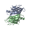
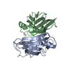
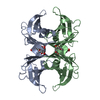
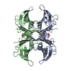
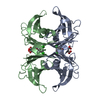
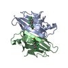
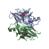
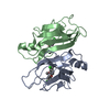
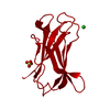
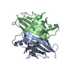
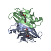
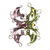
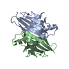
| Title | Crystal structure of transthyretin in complex with CHF5074 at neutral pH | ||||||
 Components Components | Transthyretin | ||||||
 Keywords Keywords | transport protein/inhibitor / amyloidosis / fibrillogenesis / amyloid fibrils / T3 or T4 hormone binding / Plasma / transport protein-inhibitor complex | ||||||
| Function / homology |  Function and homology information Function and homology informationRetinoid cycle disease events / thyroid hormone binding / The canonical retinoid cycle in rods (twilight vision) / Non-integrin membrane-ECM interactions / purine nucleobase metabolic process / Retinoid metabolism and transport / hormone activity / azurophil granule lumen / Amyloid fiber formation / Neutrophil degranulation ...Retinoid cycle disease events / thyroid hormone binding / The canonical retinoid cycle in rods (twilight vision) / Non-integrin membrane-ECM interactions / purine nucleobase metabolic process / Retinoid metabolism and transport / hormone activity / azurophil granule lumen / Amyloid fiber formation / Neutrophil degranulation / extracellular space / extracellular exosome / extracellular region / identical protein bindingSimilarity search - Function | ||||||
| Biological species |  Homo sapiens (human) Homo sapiens (human) | ||||||
| Method | X-RAY DIFFRACTION / SYNCHROTRON / MOLECULAR REPLACEMENT / Resolution: 1.67 Å | ||||||
 Authors Authors | Zanotti, G. / Cendron, L. / Folli, C. / Florio, P. / Imbimbo, B.P. / Berni, R. | ||||||
 Citation Citation | Journal: Febs Lett. / Year: 2013 Title: Structural evidence for native state stabilization of a conformationally labile amyloidogenic transthyretin variant by fibrillogenesis inhibitors. Authors: Zanotti, G. / Cendron, L. / Folli, C. / Florio, P. / Imbimbo, B.P. / Berni, R. #1: Journal: J.Mol.Biol. / Year: 2007Title: Acidic pH-induced conformational changes in amyloidogenic mutant transthyretin. Authors: Pasquato, N. / Berni, R. / Folli, C. / Alfieri, B. / Cendron, L. / Zanotti, G. #2: Journal: J.Biol.Chem. / Year: 2009Title: Amyloidogenic potential of transthyretin variants: insights from structural and computational analyses. Authors: Cendron, L. / Trovato, A. / Seno, F. / Folli, C. / Alfieri, B. / Zanotti, G. / Berni, R. | ||||||
| History |
|
- Structure visualization
| Structure viewer | Molecule: MolmilJmol/JSmol |
|---|
- Downloads & links
-Download
| PDBx/mmCIF format | 4i85.cif.gz | 62.6 KB | Display | PDBx/mmCIF format |
|---|---|---|---|---|
| PDB format | pdb4i85.ent.gz | 46.2 KB | Display | PDB format |
| PDBx/mmJSON format | 4i85.json.gz | Tree view | PDBx/mmJSON format | |
| Others |  Other downloads Other downloads |
-Validation report
| Arichive directory | https://data.pdbj.org/pub/pdb/validation_reports/i8/4i85ftp://data.pdbj.org/pub/pdb/validation_reports/i8/4i85 | HTTPS FTP |
|---|
-Related structure data
| Related structure data |  4i87C 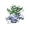 4i89C 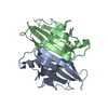 1f41S S: Starting model for refinement C: citing same article ( |
|---|---|
| Similar structure data |


-Links
 PDBj
PDBj


- Assembly
| Deposited unit | 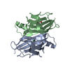
| |||||||||||||||||||||
|---|---|---|---|---|---|---|---|---|---|---|---|---|---|---|---|---|---|---|---|---|---|---|
| 1 | 
| |||||||||||||||||||||
| Unit cell |
| |||||||||||||||||||||
| Components on special symmetry positions |
|
-Components
| #1: Protein | / ATTR / Prealbumin / TBPA Mass: 13777.360 Da / Num. of mol.: 2 / Fragment: Transthyretin (unp residues 21-147) Source method: isolated from a genetically manipulated source Source: (gene. exp.) Homo sapiens (human) / Gene: PALB, TTR / Production host:  Escherichia coli (E. coli) / Strain (production host): BL21(DE3) / References: UniProt: P02766 Escherichia coli (E. coli) / Strain (production host): BL21(DE3) / References: UniProt: P02766#2: Chemical |   Mass: 325.162 Da / Num. of mol.: 2 / Source method: obtained synthetically / Formula: C16H11Cl2FO2 Mass: 325.162 Da / Num. of mol.: 2 / Source method: obtained synthetically / Formula: C16H11Cl2FO2#3: Water | ChemComp-HOH / | Water Mass: 18.015 Da / Num. of mol.: 197 / Source method: isolated from a natural source / Formula: H2O Mass: 18.015 Da / Num. of mol.: 197 / Source method: isolated from a natural source / Formula: H2O |
|---|
-Experimental details
-Experiment
| Experiment | Method: X-RAY DIFFRACTION / Number of used crystals: 1 |
|---|
- Sample preparation
| Crystal | Density Matthews: 2.08 Å3/Da / Density % sol: 40.89 % |
|---|---|
| Crystal grow | Temperature: 298 K / Method: vapor diffusion, sitting drop / pH: 7 Details: 2 M ammonium sulfate, 0.1 M KCl, 0.05 M sodium phosphate, pH 7, VAPOR DIFFUSION, SITTING DROP, temperature 298K |
-Data collection
| Diffraction | Mean temperature: 100 K |
|---|---|
| Diffraction source | Source: SYNCHROTRON / Site: ESRF  / Beamline: ID14-1 / Wavelength: 0.9334 Å / Beamline: ID14-1 / Wavelength: 0.9334 Å |
| Detector | Type: ADSC QUANTUM 210 / Detector: CCD / Date: Apr 26, 2011 |
| Radiation | Protocol: SINGLE WAVELENGTH / Monochromatic (M) / Laue (L): M / Scattering type: x-ray |
| Radiation wavelength | Wavelength: 0.9334 Å / Relative weight: 1 |
| Reflection | Resolution: 1.67→29.94 Å / Num. all: 27434 / Num. obs: 27434 / % possible obs: 99.8 % / Observed criterion σ(F): 0 / Observed criterion σ(I): 0 / Redundancy: 6.7 % / Rmerge(I) obs: 0.089 / Net I/σ(I): 15.1 |
| Reflection shell | Resolution: 1.67→1.76 Å / Redundancy: 6.3 % / Rmerge(I) obs: 0.386 / Mean I/σ(I) obs: 4.3 / Num. unique all: 3938 / % possible all: 98.8 |
- Processing
| Software |
| ||||||||||||||||||||||||||||||||||||||||||||||||||||||||||||||||||||||||||||||||||||||||||||||||||||||||||||||||||||||||||||||||||||||||||||||||||||||||||||||||||||||||||
|---|---|---|---|---|---|---|---|---|---|---|---|---|---|---|---|---|---|---|---|---|---|---|---|---|---|---|---|---|---|---|---|---|---|---|---|---|---|---|---|---|---|---|---|---|---|---|---|---|---|---|---|---|---|---|---|---|---|---|---|---|---|---|---|---|---|---|---|---|---|---|---|---|---|---|---|---|---|---|---|---|---|---|---|---|---|---|---|---|---|---|---|---|---|---|---|---|---|---|---|---|---|---|---|---|---|---|---|---|---|---|---|---|---|---|---|---|---|---|---|---|---|---|---|---|---|---|---|---|---|---|---|---|---|---|---|---|---|---|---|---|---|---|---|---|---|---|---|---|---|---|---|---|---|---|---|---|---|---|---|---|---|---|---|---|---|---|---|---|---|---|---|
| Refinement | Method to determine structure: MOLECULAR REPLACEMENT Starting model: 1F41 Resolution: 1.67→29.94 Å / Cor.coef. Fo:Fc: 0.951 / Cor.coef. Fo:Fc free: 0.922 / SU B: 2.063 / SU ML: 0.07 / Cross valid method: THROUGHOUT / ESU R: 0.106 / ESU R Free: 0.109 / Stereochemistry target values: MAXIMUM LIKELIHOOD / Details: HYDROGENS HAVE BEEN ADDED IN THE RIDING POSITIONS
| ||||||||||||||||||||||||||||||||||||||||||||||||||||||||||||||||||||||||||||||||||||||||||||||||||||||||||||||||||||||||||||||||||||||||||||||||||||||||||||||||||||||||||
| Solvent computation | Ion probe radii: 0.8 Å / Shrinkage radii: 0.8 Å / VDW probe radii: 1.4 Å / Solvent model: BABINET MODEL WITH MASK | ||||||||||||||||||||||||||||||||||||||||||||||||||||||||||||||||||||||||||||||||||||||||||||||||||||||||||||||||||||||||||||||||||||||||||||||||||||||||||||||||||||||||||
| Displacement parameters | Biso mean: 18.798 Å2
| ||||||||||||||||||||||||||||||||||||||||||||||||||||||||||||||||||||||||||||||||||||||||||||||||||||||||||||||||||||||||||||||||||||||||||||||||||||||||||||||||||||||||||
| Refinement step | Cycle: LAST / Resolution: 1.67→29.94 Å
| ||||||||||||||||||||||||||||||||||||||||||||||||||||||||||||||||||||||||||||||||||||||||||||||||||||||||||||||||||||||||||||||||||||||||||||||||||||||||||||||||||||||||||
| Refine LS restraints |
| ||||||||||||||||||||||||||||||||||||||||||||||||||||||||||||||||||||||||||||||||||||||||||||||||||||||||||||||||||||||||||||||||||||||||||||||||||||||||||||||||||||||||||
| LS refinement shell | Resolution: 1.67→1.713 Å / Total num. of bins used: 20
|
