 Movie
Movie Controller

[English] 日本語
 Yorodumi
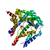
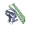
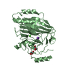
- PDB-4hwy: Trypanosoma brucei procathepsin B solved from 40 fs free-electron... -
+ Open data

- Basic information
| Entry | Database: PDB / ID: 4hwy | |||||||||
|---|---|---|---|---|---|---|---|---|---|---|
| Title | Trypanosoma brucei procathepsin B solved from 40 fs free-electron laser pulse data by serial femtosecond X-ray crystallography | |||||||||
 Components Components | Cysteine peptidase C (CPC) | |||||||||
 Keywords Keywords |  HYDROLASE / Papain fold / Lysosomal cysteine protease HYDROLASE / Papain fold / Lysosomal cysteine protease | |||||||||
| Function / homology |  Function and homology information Function and homology informationpost-transcriptional regulation of gene expression / proteolysis involved in protein catabolic process / Hydrolases; Acting on peptide bonds (peptidases); Cysteine endopeptidases / cysteine-type endopeptidase activity / extracellular spaceSimilarity search - Function | |||||||||
| Biological species |  Trypanosoma brucei brucei TREU927 (eukaryote) Trypanosoma brucei brucei TREU927 (eukaryote) | |||||||||
| Method | X-RAY DIFFRACTION / FREE ELECTRON LASER / MOLECULAR REPLACEMENT / Resolution: 2.1 Å | |||||||||
 Authors Authors | Redecke, L. / Nass, K. / DePonte, D.P. / White, T.A. / Rehders, D. / Barty, A. / Stellato, F. / Liang, M. / Barends, T.R.M. / Boutet, S. ...Redecke, L. / Nass, K. / DePonte, D.P. / White, T.A. / Rehders, D. / Barty, A. / Stellato, F. / Liang, M. / Barends, T.R.M. / Boutet, S. / Williams, G.W. / Messerschmidt, M. / Seibert, M.M. / Aquila, A. / Arnlund, D. / Bajt, S. / Barth, T. / Bogan, M.J. / Caleman, C. / Chao, T.-C. / Doak, R.B. / Fleckenstein, H. / Frank, M. / Fromme, R. / Galli, L. / Grotjohann, I. / Hunter, M.S. / Johansson, L.C. / Kassemeyer, S. / Katona, G. / Kirian, R.A. / Koopmann, R. / Kupitz, C. / Lomb, L. / Martin, A.V. / Mogk, S. / Neutze, R. / Shoemann, R.L. / Steinbrener, J. / Timneanu, N. / Wang, D. / Weierstall, U. / Zatsepin, N.A. / Spence, J.C.H. / Fromme, P. / Schlichting, I. / Duszenko, M. / Betzel, C. / Chapman, H. | |||||||||
 Citation Citation | Journal: Science / Year: 2013 Title: Natively inhibited Trypanosoma brucei cathepsin B structure determined by using an X-ray laser. Authors: Redecke, L. / Nass, K. / DePonte, D.P. / White, T.A. / Rehders, D. / Barty, A. / Stellato, F. / Liang, M. / Barends, T.R. / Boutet, S. / Williams, G.J. / Messerschmidt, M. / Seibert, M.M. / ...Authors: Redecke, L. / Nass, K. / DePonte, D.P. / White, T.A. / Rehders, D. / Barty, A. / Stellato, F. / Liang, M. / Barends, T.R. / Boutet, S. / Williams, G.J. / Messerschmidt, M. / Seibert, M.M. / Aquila, A. / Arnlund, D. / Bajt, S. / Barth, T. / Bogan, M.J. / Caleman, C. / Chao, T.C. / Doak, R.B. / Fleckenstein, H. / Frank, M. / Fromme, R. / Galli, L. / Grotjohann, I. / Hunter, M.S. / Johansson, L.C. / Kassemeyer, S. / Katona, G. / Kirian, R.A. / Koopmann, R. / Kupitz, C. / Lomb, L. / Martin, A.V. / Mogk, S. / Neutze, R. / Shoeman, R.L. / Steinbrener, J. / Timneanu, N. / Wang, D. / Weierstall, U. / Zatsepin, N.A. / Spence, J.C. / Fromme, P. / Schlichting, I. / Duszenko, M. / Betzel, C. / Chapman, H.N. | |||||||||
| History |
|
- Structure visualization
| Structure viewer | Molecule: MolmilJmol/JSmol |
|---|
- Downloads & links
-Download
| PDBx/mmCIF format | 4hwy.cif.gz | 80 KB | Display | PDBx/mmCIF format |
|---|---|---|---|---|
| PDB format | pdb4hwy.ent.gz | 57.9 KB | Display | PDB format |
| PDBx/mmJSON format | 4hwy.json.gz | Tree view | PDBx/mmJSON format | |
| Others |  Other downloads Other downloads |
-Validation report
| Arichive directory | https://data.pdbj.org/pub/pdb/validation_reports/hw/4hwyftp://data.pdbj.org/pub/pdb/validation_reports/hw/4hwy | HTTPS FTP |
|---|
-Related structure data
| Related structure data |  3morS S: Starting model for refinement |
|---|---|
| Similar structure data |


-Links
 PDBj
PDBj
- Assembly
| Deposited unit | 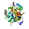
| |||||||||
|---|---|---|---|---|---|---|---|---|---|---|
| 1 |
| |||||||||
| Unit cell |
| |||||||||
| Components on special symmetry positions |
|
-Components
| #1: Protein | Mass: 37259.688 Da / Num. of mol.: 1 Source method: isolated from a genetically manipulated source Source: (gene. exp.) Trypanosoma brucei brucei TREU927 (eukaryote)Gene: Tb927.6.560 / Plasmid: pFastBac1 / Production host:   Spodoptera frugiperda (fall armyworm) / Strain (production host): SF9 Spodoptera frugiperda (fall armyworm) / Strain (production host): SF9References: UniProt: D6XHE1, Hydrolases; Acting on peptide bonds (peptidases); Cysteine endopeptidases |
|---|---|
| #2: Polysaccharide | beta-D-mannopyranose-(1-4)-2-acetamido-2-deoxy-beta-D-glucopyranose-(1-4)-2-acetamido-2-deoxy-beta- ...beta-D-mannopyranose-(1-4)-2-acetamido-2-deoxy-beta-D-glucopyranose-(1-4)-2-acetamido-2-deoxy-beta-D-glucopyranose / Mass: 586.542 Da / Num. of mol.: 1 Source method: isolated from a genetically manipulated source |
| #3: Polysaccharide | 2-acetamido-2-deoxy-beta-D-glucopyranose-(1-4)-2-acetamido-2-deoxy-beta-D-glucopyranose / Mass: 424.401 Da / Num. of mol.: 1 Source method: isolated from a genetically manipulated source |
| #4: Water | ChemComp-HOH / Water Mass: 18.015 Da / Num. of mol.: 98 / Source method: isolated from a natural source / Formula: H2O Mass: 18.015 Da / Num. of mol.: 98 / Source method: isolated from a natural source / Formula: H2O |
-Experimental details
-Experiment
| Experiment | Method: X-RAY DIFFRACTION / Number of used crystals: 293195 |
|---|
- Sample preparation
| Crystal | Density Matthews: 2.88 Å3/Da / Density % sol: 57.27 % |
|---|---|
| Crystal grow | Temperature: 310.15 K Method: crystallization in vivo within living sf9 insect cells pH: 7.4 Details: Spontaneous formation of needle-shaped microcrystals in Sf9 cells infected with recombinant baculovirus containing the gene encoding the pre-pro form of Trypanosoma brucei cathepsin B, pH 7. ...Details: Spontaneous formation of needle-shaped microcrystals in Sf9 cells infected with recombinant baculovirus containing the gene encoding the pre-pro form of Trypanosoma brucei cathepsin B, pH 7.4, Crystallization in vivo within living SF9 insect cells, temperature 310.15K |
-Data collection
| Diffraction | Mean temperature: 293 K |
|---|---|
| Diffraction source | Source: FREE ELECTRON LASER / Site: SLAC LCLS  / Beamline: CXI / Wavelength: 1.32 Å / Beamline: CXI / Wavelength: 1.32 Å |
| Detector | Type: Cornell-SLAC Pixel Array Detector / Detector: PIXEL / Date: Feb 1, 2011 |
| Radiation | Monochromator: CXI / Protocol: SINGLE WAVELENGTH / Monochromatic (M) / Laue (L): M / Scattering type: x-ray |
| Radiation wavelength | Wavelength: 1.32 Å / Relative weight: 1 |
| Reflection | Resolution: 2.1→20 Å / Num. all: 25969 / Num. obs: 25969 / % possible obs: 100 % / Observed criterion σ(F): 0 / Observed criterion σ(I): 0 |
| Reflection shell | Resolution: 2.1→2.175 Å / % possible all: 100 |
- Processing
| Software |
| ||||||||||||||||||||||||||||||||||||||||||||||||||||||||||||||||||||||||||||||||||||||||||||||||||||||||||||||||||||||||||||||||||||||||||||||||||||||||||||||||||||||||||
|---|---|---|---|---|---|---|---|---|---|---|---|---|---|---|---|---|---|---|---|---|---|---|---|---|---|---|---|---|---|---|---|---|---|---|---|---|---|---|---|---|---|---|---|---|---|---|---|---|---|---|---|---|---|---|---|---|---|---|---|---|---|---|---|---|---|---|---|---|---|---|---|---|---|---|---|---|---|---|---|---|---|---|---|---|---|---|---|---|---|---|---|---|---|---|---|---|---|---|---|---|---|---|---|---|---|---|---|---|---|---|---|---|---|---|---|---|---|---|---|---|---|---|---|---|---|---|---|---|---|---|---|---|---|---|---|---|---|---|---|---|---|---|---|---|---|---|---|---|---|---|---|---|---|---|---|---|---|---|---|---|---|---|---|---|---|---|---|---|---|---|---|
| Refinement | Method to determine structure: MOLECULAR REPLACEMENT Starting model: PDB entry 3MOR Resolution: 2.1→88.67 Å / Cor.coef. Fo:Fc: 0.964 / Cor.coef. Fo:Fc free: 0.957 / SU B: 4.531 / SU ML: 0.113 / Cross valid method: THROUGHOUT / ESU R: 0.161 / ESU R Free: 0.145 / Stereochemistry target values: MAXIMUM LIKELIHOOD / Details: HYDROGENS HAVE BEEN USED IF PRESENT IN THE INPUT
| ||||||||||||||||||||||||||||||||||||||||||||||||||||||||||||||||||||||||||||||||||||||||||||||||||||||||||||||||||||||||||||||||||||||||||||||||||||||||||||||||||||||||||
| Solvent computation | Ion probe radii: 0.8 Å / Shrinkage radii: 0.8 Å / VDW probe radii: 1.2 Å / Solvent model: MASK | ||||||||||||||||||||||||||||||||||||||||||||||||||||||||||||||||||||||||||||||||||||||||||||||||||||||||||||||||||||||||||||||||||||||||||||||||||||||||||||||||||||||||||
| Displacement parameters | Biso mean: 46.244 Å2
| ||||||||||||||||||||||||||||||||||||||||||||||||||||||||||||||||||||||||||||||||||||||||||||||||||||||||||||||||||||||||||||||||||||||||||||||||||||||||||||||||||||||||||
| Refinement step | Cycle: LAST / Resolution: 2.1→88.67 Å
| ||||||||||||||||||||||||||||||||||||||||||||||||||||||||||||||||||||||||||||||||||||||||||||||||||||||||||||||||||||||||||||||||||||||||||||||||||||||||||||||||||||||||||
| Refine LS restraints |
| ||||||||||||||||||||||||||||||||||||||||||||||||||||||||||||||||||||||||||||||||||||||||||||||||||||||||||||||||||||||||||||||||||||||||||||||||||||||||||||||||||||||||||
| LS refinement shell | Resolution: 2.1→2.155 Å / Total num. of bins used: 20
|
