 Movie
Movie Controller

[English] 日本語
 Yorodumi
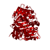
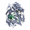
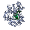
- PDB-4gvr: X-ray structure of the Archaeoglobus fulgidus methenyl-tetrahydro... -
+ Open data

- Basic information
| Entry | Database: PDB / ID: 4gvr | ||||||
|---|---|---|---|---|---|---|---|
| Title | X-ray structure of the Archaeoglobus fulgidus methenyl-tetrahydromethanopterin cyclohydrolase | ||||||
 Components Components | Methenyltetrahydromethanopterin cyclohydrolase | ||||||
 Keywords Keywords | HYDROLASE / methenyl-tetrahydromethanopterin / N5-formyl-tetrahydromethanopterin | ||||||
| Function / homology |  Function and homology informationmethenyltetrahydromethanopterin cyclohydrolase / methenyltetrahydromethanopterin cyclohydrolase activity / lactate oxidation / one-carbon metabolic process / cytoplasm Function and homology informationmethenyltetrahydromethanopterin cyclohydrolase / methenyltetrahydromethanopterin cyclohydrolase activity / lactate oxidation / one-carbon metabolic process / cytoplasmSimilarity search - Function | ||||||
| Biological species |   Archaeoglobus fulgidus (archaea) Archaeoglobus fulgidus (archaea) | ||||||
| Method | X-RAY DIFFRACTION / SYNCHROTRON / MOLECULAR REPLACEMENT / Resolution: 1.52 Å | ||||||
 Authors Authors | Upadhyay, V. / Demmer, U. / Warkentin, E. / Moll, J. / Shima, S. / Ermler, U. | ||||||
 Citation Citation | Journal: Biochemistry / Year: 2012 Title: Structure and catalytic mechanism of N(5),N(10)-methenyl-tetrahydromethanopterin cyclohydrolase. Authors: Upadhyay, V. / Demmer, U. / Warkentin, E. / Moll, J. / Shima, S. / Ermler, U. | ||||||
| History |
|
- Structure visualization
| Structure viewer | Molecule: MolmilJmol/JSmol |
|---|
- Downloads & links
-Download
| PDBx/mmCIF format | 4gvr.cif.gz | 410.9 KB | Display | PDBx/mmCIF format |
|---|---|---|---|---|
| PDB format | pdb4gvr.ent.gz | 339 KB | Display | PDB format |
| PDBx/mmJSON format | 4gvr.json.gz | Tree view | PDBx/mmJSON format | |
| Others |  Other downloads Other downloads |
-Validation report
| Arichive directory | https://data.pdbj.org/pub/pdb/validation_reports/gv/4gvrftp://data.pdbj.org/pub/pdb/validation_reports/gv/4gvr | HTTPS FTP |
|---|
-Related structure data
| Related structure data |  4gvqC 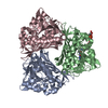 4gvsC  1qlmS C: citing same article ( S: Starting model for refinement |
|---|---|
| Similar structure data |


-Links
 PDBj
PDBj- Assembly
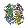
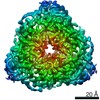
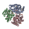
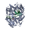
| Deposited unit | 
| ||||||||||||||||||||||||||||||||||||||||||||||||||||
|---|---|---|---|---|---|---|---|---|---|---|---|---|---|---|---|---|---|---|---|---|---|---|---|---|---|---|---|---|---|---|---|---|---|---|---|---|---|---|---|---|---|---|---|---|---|---|---|---|---|---|---|---|---|
| 1 |
| ||||||||||||||||||||||||||||||||||||||||||||||||||||
| Unit cell |
| ||||||||||||||||||||||||||||||||||||||||||||||||||||
| Noncrystallographic symmetry (NCS) | NCS domain:
NCS domain segments:
NCS oper:
|
-Components
| #1: Protein | / Methenyl-H4MPT cyclohydrolase Mass: 34879.637 Da / Num. of mol.: 3 / Mutation: E186Q Source method: isolated from a genetically manipulated source Source: (gene. exp.) Archaeoglobus fulgidus (archaea) / Strain: DSM 4304 / Gene: AF_1935, mch / Plasmid: pET-24a(+) / Production host:  Escherichia coli (E. coli) / Strain (production host): Rosetta (DE3) pLysS Escherichia coli (E. coli) / Strain (production host): Rosetta (DE3) pLysSReferences: UniProt: O28344, methenyltetrahydromethanopterin cyclohydrolase#2: Water | ChemComp-HOH / | Water Mass: 18.015 Da / Num. of mol.: 740 / Source method: isolated from a natural source / Formula: H2O Mass: 18.015 Da / Num. of mol.: 740 / Source method: isolated from a natural source / Formula: H2O |
|---|
-Experimental details
-Experiment
| Experiment | Method: X-RAY DIFFRACTION / Number of used crystals: 1 |
|---|
- Sample preparation
| Crystal | Density Matthews: 2.71 Å3/Da / Density % sol: 54.63 % |
|---|---|
| Crystal grow | Temperature: 291 K / Method: vapor diffusion, hanging drop / pH: 5.6 Details: 10% (w/v) PEG 4000, 10% (v/v) Isopropanol, 0.1M Sodium acetate pH 5.6, VAPOR DIFFUSION, HANGING DROP, temperature 291K |
-Data collection
| Diffraction source | Source: SYNCHROTRON / Site: SLS  / Beamline: X10SA / Wavelength: 1.0001 Å / Beamline: X10SA / Wavelength: 1.0001 Å |
|---|---|
| Detector | Date: Dec 8, 2011 |
| Radiation | Protocol: SINGLE WAVELENGTH / Monochromatic (M) / Laue (L): M / Scattering type: x-ray |
| Radiation wavelength | Wavelength: 1.0001 Å / Relative weight: 1 |
| Reflection | Resolution: 1.52→48.17 Å / Num. all: 170753 / Num. obs: 164493 |
- Processing
| Software |
| ||||||||||||||||||||||||||||||||||||||||||||||||||||||||||||||||||||||||||||||||||||||||||||||||||||||||||||||||||||||||||||||||||||||||||||||||||||||||||||||||||||||||||
|---|---|---|---|---|---|---|---|---|---|---|---|---|---|---|---|---|---|---|---|---|---|---|---|---|---|---|---|---|---|---|---|---|---|---|---|---|---|---|---|---|---|---|---|---|---|---|---|---|---|---|---|---|---|---|---|---|---|---|---|---|---|---|---|---|---|---|---|---|---|---|---|---|---|---|---|---|---|---|---|---|---|---|---|---|---|---|---|---|---|---|---|---|---|---|---|---|---|---|---|---|---|---|---|---|---|---|---|---|---|---|---|---|---|---|---|---|---|---|---|---|---|---|---|---|---|---|---|---|---|---|---|---|---|---|---|---|---|---|---|---|---|---|---|---|---|---|---|---|---|---|---|---|---|---|---|---|---|---|---|---|---|---|---|---|---|---|---|---|---|---|---|
| Refinement | Method to determine structure: MOLECULAR REPLACEMENT Starting model: 1QLM Resolution: 1.52→48.17 Å / Cor.coef. Fo:Fc: 0.971 / Cor.coef. Fo:Fc free: 0.944 / SU B: 4.941 / SU ML: 0.075 / Cross valid method: THROUGHOUT / ESU R: 0.082 / ESU R Free: 0.083 / Stereochemistry target values: MAXIMUM LIKELIHOOD / Details: HYDROGENS HAVE BEEN USED IF PRESENT IN THE INPUT
| ||||||||||||||||||||||||||||||||||||||||||||||||||||||||||||||||||||||||||||||||||||||||||||||||||||||||||||||||||||||||||||||||||||||||||||||||||||||||||||||||||||||||||
| Solvent computation | Ion probe radii: 0.8 Å / Shrinkage radii: 0.8 Å / VDW probe radii: 1.2 Å / Solvent model: MASK | ||||||||||||||||||||||||||||||||||||||||||||||||||||||||||||||||||||||||||||||||||||||||||||||||||||||||||||||||||||||||||||||||||||||||||||||||||||||||||||||||||||||||||
| Displacement parameters | Biso mean: 25.003 Å2
| ||||||||||||||||||||||||||||||||||||||||||||||||||||||||||||||||||||||||||||||||||||||||||||||||||||||||||||||||||||||||||||||||||||||||||||||||||||||||||||||||||||||||||
| Refinement step | Cycle: LAST / Resolution: 1.52→48.17 Å
| ||||||||||||||||||||||||||||||||||||||||||||||||||||||||||||||||||||||||||||||||||||||||||||||||||||||||||||||||||||||||||||||||||||||||||||||||||||||||||||||||||||||||||
| Refine LS restraints |
| ||||||||||||||||||||||||||||||||||||||||||||||||||||||||||||||||||||||||||||||||||||||||||||||||||||||||||||||||||||||||||||||||||||||||||||||||||||||||||||||||||||||||||
| Refine LS restraints NCS | Ens-ID: 1 / Number: 2347 / Refine-ID: X-RAY DIFFRACTION
| ||||||||||||||||||||||||||||||||||||||||||||||||||||||||||||||||||||||||||||||||||||||||||||||||||||||||||||||||||||||||||||||||||||||||||||||||||||||||||||||||||||||||||
| LS refinement shell | Resolution: 1.52→1.559 Å / Total num. of bins used: 20
|
