 Movie
Movie Controller


+ Open data

- Basic information
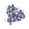
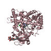
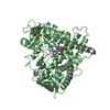
| Entry | Database: PDB / ID: 4gqs | ||||||
|---|---|---|---|---|---|---|---|
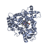
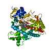
| Title | Structure of Human Microsomal Cytochrome P450 (CYP) 2C19 | ||||||
 Components Components | Cytochrome P450 2C19 | ||||||
 Keywords Keywords |  OXIDOREDUCTASE / monooxygenase / drug metabolizing enzyme / heme protein OXIDOREDUCTASE / monooxygenase / drug metabolizing enzyme / heme protein | ||||||
| Function / homology |  Function and homology information Function and homology informationfenbendazole monooxygenase (4'-hydroxylating) / xenobiotic catabolic process => GO:0042178 / xenobiotic metabolic process => GO:0006805 / (S)-limonene 6-monooxygenase / (S)-limonene 7-monooxygenase / (R)-limonene 6-monooxygenase / (S)-limonene 6-monooxygenase activity / (S)-limonene 7-monooxygenase activity / (R)-limonene 6-monooxygenase activity / organic acid metabolic process ...fenbendazole monooxygenase (4'-hydroxylating) / xenobiotic catabolic process => GO:0042178 / xenobiotic metabolic process => GO:0006805 / (S)-limonene 6-monooxygenase / (S)-limonene 7-monooxygenase / (R)-limonene 6-monooxygenase / (S)-limonene 6-monooxygenase activity / (S)-limonene 7-monooxygenase activity / (R)-limonene 6-monooxygenase activity / organic acid metabolic process / Synthesis of (16-20)-hydroxyeicosatetraenoic acids (HETE) / omega-hydroxylase P450 pathway / arachidonic acid epoxygenase activity / CYP2E1 reactions / epoxygenase P450 pathway / : / heterocycle metabolic process / Synthesis of epoxy (EET) and dihydroxyeicosatrienoic acids (DHET) / monoterpenoid metabolic process / Xenobiotics / steroid hydroxylase activity / oxidoreductase activity, acting on paired donors, with incorporation or reduction of molecular oxygen, reduced flavin or flavoprotein as one donor, and incorporation of one atom of oxygen / unspecific monooxygenase / aromatase activity / steroid metabolic process / xenobiotic metabolic process / monooxygenase activity / oxygen binding / oxidoreductase activity / iron ion binding / intracellular membrane-bounded organelle / heme binding / endoplasmic reticulum membrane / enzyme binding / cytoplasmSimilarity search - Function | ||||||
| Biological species |  Homo sapiens (human) Homo sapiens (human) | ||||||
| Method | X-RAY DIFFRACTION / SYNCHROTRON / MOLECULAR REPLACEMENT / Resolution: 2.87 Å | ||||||
 Authors Authors | Reynald, R.L. / Sansen, S. / Stout, C.D. / Johnson, E.F. | ||||||
 Citation Citation | Journal: J.Biol.Chem. / Year: 2012 Title: Structural Characterization of Human Cytochrome P450 2C19: ACTIVE SITE DIFFERENCES BETWEEN P450s 2C8, 2C9, AND 2C19. Authors: Reynald, R.L. / Sansen, S. / Stout, C.D. / Johnson, E.F. | ||||||
| History |
|
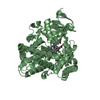
- Structure visualization
| Structure viewer | Molecule: MolmilJmol/JSmol |
|---|
- Downloads & links
-Download
| PDBx/mmCIF format | 4gqs.cif.gz | 372.6 KB | Display | PDBx/mmCIF format |
|---|---|---|---|---|
| PDB format | pdb4gqs.ent.gz | 305.6 KB | Display | PDB format |
| PDBx/mmJSON format | 4gqs.json.gz | Tree view | PDBx/mmJSON format | |
| Others |  Other downloads Other downloads |
-Validation report
| Arichive directory | https://data.pdbj.org/pub/pdb/validation_reports/gq/4gqsftp://data.pdbj.org/pub/pdb/validation_reports/gq/4gqs | HTTPS FTP |
|---|
-Related structure data
| Related structure data |  1r9oS S: Starting model for refinement |
|---|---|
| Similar structure data |


-Links
 PDBj
PDBj


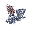
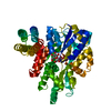
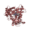
- Assembly
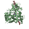
| Deposited unit | 
| ||||||||
|---|---|---|---|---|---|---|---|---|---|
| 1 | 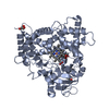
| ||||||||
| 2 | 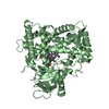
| ||||||||
| 3 | 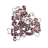
| ||||||||
| 4 | 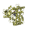
| ||||||||
| Unit cell |
|
-Components
| #1: Protein | Mass: 54581.000 Da / Num. of mol.: 4 / Fragment: CATALYTIC DOMAIN (UNP Residues 21-490) Source method: isolated from a genetically manipulated source Source: (gene. exp.) Homo sapiens (human) / Strain: ALLELIC VARIANT 1B / Gene: CYP2C19 / Plasmid: PCW0RI / Production host:  Escherichia coli (E. coli) / Strain (production host): DH5alpha Escherichia coli (E. coli) / Strain (production host): DH5alphaReferences: UniProt: P33261, Oxidoreductases; Acting on paired donors, with incorporation or reduction of molecular oxygen; With NADH or NADPH as one donor, and incorporation of one atom of oxygen ...References: UniProt: P33261, Oxidoreductases; Acting on paired donors, with incorporation or reduction of molecular oxygen; With NADH or NADPH as one donor, and incorporation of one atom of oxygen into the other donor, EC: 1.14.13.80, EC: 1.14.13.48, EC: 1.14.13.49#2: Chemical | ChemComp-HEM / Heme B  Mass: 616.487 Da / Num. of mol.: 4 / Source method: obtained synthetically / Formula: C34H32FeN4O4 Mass: 616.487 Da / Num. of mol.: 4 / Source method: obtained synthetically / Formula: C34H32FeN4O4#3: Chemical | ChemComp-0XV / ( 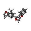  Mass: 280.318 Da / Num. of mol.: 4 / Source method: obtained synthetically / Formula: C18H16O3 Mass: 280.318 Da / Num. of mol.: 4 / Source method: obtained synthetically / Formula: C18H16O3#4: Chemical | Glycerol  Mass: 92.094 Da / Num. of mol.: 3 / Source method: obtained synthetically / Formula: C3H8O3 Mass: 92.094 Da / Num. of mol.: 3 / Source method: obtained synthetically / Formula: C3H8O3#5: Water | ChemComp-HOH / | Water Mass: 18.015 Da / Num. of mol.: 50 / Source method: isolated from a natural source / Formula: H2O Mass: 18.015 Da / Num. of mol.: 50 / Source method: isolated from a natural source / Formula: H2O |
|---|
-Experimental details
-Experiment
| Experiment | Method: X-RAY DIFFRACTION / Number of used crystals: 1 |
|---|
- Sample preparation
| Crystal | Density Matthews: 2.51 Å3/Da / Density % sol: 51.06 % |
|---|---|
| Crystal grow | Temperature: 297 K / Method: vapor diffusion, hanging drop / pH: 7.5 Details: 20% PEG 2000, 10% isopropanol, 0.1M HEPES, pH 7.5, VAPOR DIFFUSION, HANGING DROP, temperature 297K |
-Data collection
| Diffraction | Mean temperature: 90 K |
|---|---|
| Diffraction source | Source: SYNCHROTRON / Site: SSRL  / Beamline: BL11-1 / Wavelength: 0.92014 Å / Beamline: BL11-1 / Wavelength: 0.92014 Å |
| Detector | Type: ADSC QUANTUM 315 / Detector: CCD |
| Radiation | Protocol: SINGLE WAVELENGTH / Monochromatic (M) / Laue (L): M / Scattering type: x-ray |
| Radiation wavelength | Wavelength: 0.92014 Å / Relative weight: 1 |
| Reflection | Resolution: 2.87→79.6 Å / Num. all: 50609 / Num. obs: 50609 / % possible obs: 100 % / Observed criterion σ(F): 0 / Redundancy: 11.8 % / Biso Wilson estimate: 76.5 Å2 / Rmerge(I) obs: 0.087 / Net I/σ(I): 3.4 |
| Reflection shell | Resolution: 2.87→3.03 Å / Redundancy: 12 % / Rmerge(I) obs: 0.498 / Mean I/σ(I) obs: 1.6 / Num. unique all: 7297 / % possible all: 100 |
- Processing
| Software |
| ||||||||||||||||||||||||||||||||||||||||||||||||||||||||||||||||||||||||||||||||
|---|---|---|---|---|---|---|---|---|---|---|---|---|---|---|---|---|---|---|---|---|---|---|---|---|---|---|---|---|---|---|---|---|---|---|---|---|---|---|---|---|---|---|---|---|---|---|---|---|---|---|---|---|---|---|---|---|---|---|---|---|---|---|---|---|---|---|---|---|---|---|---|---|---|---|---|---|---|---|---|---|---|
| Refinement | Method to determine structure: MOLECULAR REPLACEMENT Starting model: PDB ENTRY 1R9O Resolution: 2.87→79.6 Å / Rfactor Rfree error: 0.006 / Data cutoff high absF: 8638772.65 / Data cutoff low absF: 0 / Isotropic thermal model: RESTRAINED / Cross valid method: THROUGHOUT / σ(F): 0 / Stereochemistry target values: Engh & Huber Details: FLAT BULK SOLVENT MODEL USED KSOL: 0.36 BSOL: 83.8113
| ||||||||||||||||||||||||||||||||||||||||||||||||||||||||||||||||||||||||||||||||
| Solvent computation | Solvent model: FLAT MODEL / Bsol: 83.8113 Å2 / ksol: 0.36 e/Å3 | ||||||||||||||||||||||||||||||||||||||||||||||||||||||||||||||||||||||||||||||||
| Displacement parameters | Biso mean: 85.9 Å2
| ||||||||||||||||||||||||||||||||||||||||||||||||||||||||||||||||||||||||||||||||
| Refine analyze |
| ||||||||||||||||||||||||||||||||||||||||||||||||||||||||||||||||||||||||||||||||
| Refinement step | Cycle: LAST / Resolution: 2.87→79.6 Å
| ||||||||||||||||||||||||||||||||||||||||||||||||||||||||||||||||||||||||||||||||
| Refine LS restraints |
| ||||||||||||||||||||||||||||||||||||||||||||||||||||||||||||||||||||||||||||||||
| Refine LS restraints NCS | NCS model details: NONE | ||||||||||||||||||||||||||||||||||||||||||||||||||||||||||||||||||||||||||||||||
| LS refinement shell | Resolution: 2.87→2.97 Å / Rfactor Rfree error: 0.027 / Total num. of bins used: 10
| ||||||||||||||||||||||||||||||||||||||||||||||||||||||||||||||||||||||||||||||||
| Xplor file |
|
