 Movie
Movie Controller

[English] 日本語
 Yorodumi
- PDB-4glr: Structure of the anti-ptau Fab (pT231/pS235_1) in complex with ph... -
+ Open data

- Basic information
| Entry | Database: PDB / ID: 4glr | ||||||
|---|---|---|---|---|---|---|---|
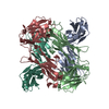
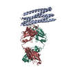
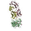
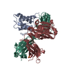
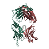
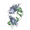
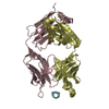
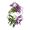
| Title | Structure of the anti-ptau Fab (pT231/pS235_1) in complex with phosphoepitope pT231/pS235 | ||||||
 Components Components |
| ||||||
 Keywords Keywords |  IMMUNE SYSTEM / IgG1 Fab / anti-ptau antibody / phosphorylated tau peptide / phosphorylation IMMUNE SYSTEM / IgG1 Fab / anti-ptau antibody / phosphorylated tau peptide / phosphorylation | ||||||
| Function / homology |  Function and homology information Function and homology informationplus-end-directed organelle transport along microtubule / axonal transport / histone-dependent DNA binding / neurofibrillary tangle assembly / positive regulation of diacylglycerol kinase activity / negative regulation of establishment of protein localization to mitochondrion / neurofibrillary tangle / positive regulation of protein localization to synapse / microtubule lateral binding / tubulin complex ...plus-end-directed organelle transport along microtubule / axonal transport / histone-dependent DNA binding / neurofibrillary tangle assembly / positive regulation of diacylglycerol kinase activity / negative regulation of establishment of protein localization to mitochondrion / neurofibrillary tangle / positive regulation of protein localization to synapse / microtubule lateral binding / tubulin complex / phosphatidylinositol bisphosphate binding / main axon / regulation of long-term synaptic depression / negative regulation of kinase activity / negative regulation of tubulin deacetylation / generation of neurons / regulation of chromosome organization / positive regulation of protein localization / rRNA metabolic process / internal protein amino acid acetylation / regulation of mitochondrial fission / lipoprotein particle binding / intracellular distribution of mitochondria / axonal transport of mitochondrion / axon development / central nervous system neuron development / regulation of microtubule polymerization / microtubule polymerization / minor groove of adenine-thymine-rich DNA binding / negative regulation of mitochondrial membrane potential / dynactin binding / glial cell projection / apolipoprotein binding / protein polymerization / negative regulation of mitochondrial fission / axolemma / Caspase-mediated cleavage of cytoskeletal proteins / regulation of microtubule polymerization or depolymerization / positive regulation of axon extension / supramolecular fiber organization / Activation of AMPK downstream of NMDARs / cytoplasmic microtubule organization / regulation of microtubule cytoskeleton organization / stress granule assembly / regulation of cellular response to heat / regulation of calcium-mediated signaling / axon cytoplasm / positive regulation of microtubule polymerization / cellular response to brain-derived neurotrophic factor stimulus / somatodendritic compartment / synapse assembly / phosphatidylinositol binding / nuclear periphery / cellular response to nerve growth factor stimulus / positive regulation of superoxide anion generation / protein phosphatase 2A binding / regulation of autophagy / astrocyte activation / synapse organization / microglial cell activation / response to lead ion / Hsp90 protein binding / regulation of synaptic plasticity / PKR-mediated signaling / protein homooligomerization / cytoplasmic ribonucleoprotein granule / memory / microtubule cytoskeleton organization / SH3 domain binding / cellular response to reactive oxygen species / neuron projection development / activation of cysteine-type endopeptidase activity involved in apoptotic process / microtubule cytoskeleton / protein-macromolecule adaptor activity / single-stranded DNA binding / cell-cell signaling / cellular response to heat / cell body / actin binding / growth cone / protein-folding chaperone binding / double-stranded DNA binding / microtubule binding / microtubule / amyloid fibril formation / sequence-specific DNA binding / dendritic spine / learning or memory / neuron projection / nuclear speck / membrane raft / axon / negative regulation of gene expression / dendrite / neuronal cell body / DNA damage response / protein kinase binding / enzyme binding / mitochondrion / DNA bindingSimilarity search - Function | ||||||
| Biological species |  Gallus gallus (chicken) Gallus gallus (chicken) Homo sapiens (human) Homo sapiens (human) | ||||||
| Method | X-RAY DIFFRACTION / SYNCHROTRON / MOLECULAR REPLACEMENT / Resolution: 1.9 Å | ||||||
 Authors Authors | Tu, C. / Mosyak, L. / Bard, J. | ||||||
 Citation Citation | Journal: J.Biol.Chem. / Year: 2012 Title: An Ultra-specific Avian Antibody to Phosphorylated Tau Protein Reveals a Unique Mechanism for Phosphoepitope Recognition. Authors: Shih, H.H. / Tu, C. / Cao, W. / Klein, A. / Ramsey, R. / Fennell, B.J. / Lambert, M. / Ni Shuilleabhain, D. / Autin, B. / Kouranova, E. / Laxmanan, S. / Braithwaite, S. / Wu, L. / Ait-Zahra, ...Authors: Shih, H.H. / Tu, C. / Cao, W. / Klein, A. / Ramsey, R. / Fennell, B.J. / Lambert, M. / Ni Shuilleabhain, D. / Autin, B. / Kouranova, E. / Laxmanan, S. / Braithwaite, S. / Wu, L. / Ait-Zahra, M. / Milici, A.J. / Dumin, J.A. / Lavallie, E.R. / Arai, M. / Corcoran, C. / Paulsen, J.E. / Gill, D. / Cunningham, O. / Bard, J. / Mosyak, L. / Finlay, W.J. | ||||||
| History |
|
- Structure visualization
| Structure viewer | Molecule: MolmilJmol/JSmol |
|---|
- Downloads & links
-Download
| PDBx/mmCIF format | 4glr.cif.gz | 352.9 KB | Display | PDBx/mmCIF format |
|---|---|---|---|---|
| PDB format | pdb4glr.ent.gz | 286.5 KB | Display | PDB format |
| PDBx/mmJSON format | 4glr.json.gz | Tree view | PDBx/mmJSON format | |
| Others |  Other downloads Other downloads |
-Validation report
| Arichive directory | https://data.pdbj.org/pub/pdb/validation_reports/gl/4glrftp://data.pdbj.org/pub/pdb/validation_reports/gl/4glr | HTTPS FTP |
|---|
-Related structure data


-Links
 PDBj
PDBj


- Assembly
| Deposited unit | 
| ||||||||
|---|---|---|---|---|---|---|---|---|---|
| 1 | 
| ||||||||
| 2 | 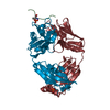
| ||||||||
| Unit cell |
|
-Components
| #1: Protein/peptide | Mass: 2048.260 Da / Num. of mol.: 2 / Fragment: phospho-peptide / Source method: obtained synthetically Details: This sequence occurs naturally in humans, except the C-terminus Cysteine Source: (synth.) Homo sapiens (human) / References: UniProt: P10636*PLUS#2: Antibody | Mass: 23609.414 Da / Num. of mol.: 2 / Fragment: heavy chain Source method: isolated from a genetically manipulated source Source: (gene. exp.) Gallus gallus (chicken) / Production host:   Cricetulus griseus (Chinese hamster) Cricetulus griseus (Chinese hamster)#3: Antibody | Mass: 22175.260 Da / Num. of mol.: 2 / Fragment: light chain Source method: isolated from a genetically manipulated source Source: (gene. exp.) Gallus gallus (chicken) / Production host: Cricetulus griseus (Chinese hamster)#4: Chemical | Phosphate  Mass: 94.971 Da / Num. of mol.: 2 / Source method: obtained synthetically / Formula: PO4 Mass: 94.971 Da / Num. of mol.: 2 / Source method: obtained synthetically / Formula: PO4#5: Water | ChemComp-HOH / | Water Mass: 18.015 Da / Num. of mol.: 715 / Source method: isolated from a natural source / Formula: H2O Mass: 18.015 Da / Num. of mol.: 715 / Source method: isolated from a natural source / Formula: H2O |
|---|
-Experimental details
-Experiment
| Experiment | Method: X-RAY DIFFRACTION / Number of used crystals: 1 |
|---|
- Sample preparation
| Crystal | Density Matthews: 2.71 Å3/Da / Density % sol: 54.66 % |
|---|---|
| Crystal grow | Temperature: 291 K / Method: vapor diffusion, sitting drop / pH: 7 Details: 200 mM NH4H2PO4, 40% MPD, pH 7.0, VAPOR DIFFUSION, SITTING DROP, temperature 291K |
-Data collection
| Diffraction | Mean temperature: 200 K |
|---|---|
| Diffraction source | Source: SYNCHROTRON / Site: APS  / Beamline: 17-ID / Wavelength: 1 Å / Beamline: 17-ID / Wavelength: 1 Å |
| Detector | Type: DECTRIS PILATUS 6M / Detector: PIXEL / Date: Aug 23, 2011 / Details: mirrors |
| Radiation | Monochromator: Si 111 / Protocol: SINGLE WAVELENGTH / Monochromatic (M) / Laue (L): M / Scattering type: x-ray |
| Radiation wavelength | Wavelength: 1 Å / Relative weight: 1 |
| Reflection | Resolution: 1.9→50 Å / Num. obs: 81511 / % possible obs: 97.9 % / Observed criterion σ(F): 1 / Observed criterion σ(I): 1 / Redundancy: 10 % / Biso Wilson estimate: 31.54 Å2 / Rmerge(I) obs: 0.059 / Net I/σ(I): 22.6 |
| Reflection shell | Resolution: 1.9→2 Å / Redundancy: 10.3 % / Rmerge(I) obs: 0.512 / Mean I/σ(I) obs: 4.1 / Num. unique all: 11379 / % possible all: 94.7 |
- Processing
| Software |
| |||||||||||||||||||||||||||||||||||||||||||||||||||||||||||||||||||||||||||||||||||||||||||||||||||||||||||||||||||||||||||||
|---|---|---|---|---|---|---|---|---|---|---|---|---|---|---|---|---|---|---|---|---|---|---|---|---|---|---|---|---|---|---|---|---|---|---|---|---|---|---|---|---|---|---|---|---|---|---|---|---|---|---|---|---|---|---|---|---|---|---|---|---|---|---|---|---|---|---|---|---|---|---|---|---|---|---|---|---|---|---|---|---|---|---|---|---|---|---|---|---|---|---|---|---|---|---|---|---|---|---|---|---|---|---|---|---|---|---|---|---|---|---|---|---|---|---|---|---|---|---|---|---|---|---|---|---|---|---|
| Refinement | Method to determine structure: MOLECULAR REPLACEMENT Starting model: PDB entries 3GJE, 3MA9 Resolution: 1.9→50 Å / Cor.coef. Fo:Fc: 0.9426 / Cor.coef. Fo:Fc free: 0.9302 / SU R Cruickshank DPI: 0.131 / Isotropic thermal model: anisotropic / Cross valid method: THROUGHOUT / σ(F): 0 / σ(I): 2 / Stereochemistry target values: Engh & Huber
| |||||||||||||||||||||||||||||||||||||||||||||||||||||||||||||||||||||||||||||||||||||||||||||||||||||||||||||||||||||||||||||
| Displacement parameters | Biso mean: 40.85 Å2
| |||||||||||||||||||||||||||||||||||||||||||||||||||||||||||||||||||||||||||||||||||||||||||||||||||||||||||||||||||||||||||||
| Refine analyze | Luzzati coordinate error obs: 0.258 Å | |||||||||||||||||||||||||||||||||||||||||||||||||||||||||||||||||||||||||||||||||||||||||||||||||||||||||||||||||||||||||||||
| Refinement step | Cycle: LAST / Resolution: 1.9→50 Å
| |||||||||||||||||||||||||||||||||||||||||||||||||||||||||||||||||||||||||||||||||||||||||||||||||||||||||||||||||||||||||||||
| Refine LS restraints |
| |||||||||||||||||||||||||||||||||||||||||||||||||||||||||||||||||||||||||||||||||||||||||||||||||||||||||||||||||||||||||||||
| LS refinement shell | Resolution: 1.9→1.95 Å / Total num. of bins used: 20
| |||||||||||||||||||||||||||||||||||||||||||||||||||||||||||||||||||||||||||||||||||||||||||||||||||||||||||||||||||||||||||||
| Refinement TLS params. | Method: refined / Refine-ID: X-RAY DIFFRACTION
| |||||||||||||||||||||||||||||||||||||||||||||||||||||||||||||||||||||||||||||||||||||||||||||||||||||||||||||||||||||||||||||
| Refinement TLS group |
|
