[English] 日本語
Yorodumi
- PDB-4gli: Crystal Structure of Human SMN YG-Dimer -

+
Open data


ID or keywords:

Loading...

-
Basic information

Entry
Database: PDB / ID: 4gli
TitleCrystal Structure of Human SMN YG-Dimer
ComponentsMaltose-binding periplasmic protein, Survival motor neuron protein chimera
KeywordsSUGAR BINDING PROTEIN / SPLICING / Soluble Glycine Zipper
Function / homology
Function and homology information


Gemini of coiled bodies / SMN complex / SMN-Sm protein complex / spliceosomal complex assembly / detection of maltose stimulus / maltose binding / maltose transport complex / maltose transport / maltodextrin transmembrane transport / carbohydrate transmembrane transporter activity ...Gemini of coiled bodies / SMN complex / SMN-Sm protein complex / spliceosomal complex assembly / detection of maltose stimulus / maltose binding / maltose transport complex / maltose transport / maltodextrin transmembrane transport / carbohydrate transmembrane transporter activity / ATP-binding cassette (ABC) transporter complex, substrate-binding subunit-containing / carbohydrate transport / Cajal body / spliceosomal snRNP assembly / ATP-binding cassette (ABC) transporter complex / cell chemotaxis / DNA-templated transcription termination / cytoplasmic ribonucleoprotein granule / Z disc / snRNP Assembly / nervous system development / outer membrane-bounded periplasmic space / SARS-CoV-2 modulates host translation machinery / perikaryon / periplasmic space / nuclear body / neuron projection / axon / DNA damage response / RNA binding / nucleoplasm / membrane / identical protein binding / nucleus / cytosol / cytoplasm
Similarity search - Function
SMN complex subunit Smn1 / : / Survival Motor Neuron, YG-box / Survival Motor Neuron, Gemin2-binding domain / : / Survival motor neuron, Tudor domain / Survival motor neuron protein (SMN), Tudor domain / Tudor domain profile. / Tudor domain / Tudor domain ...SMN complex subunit Smn1 / : / Survival Motor Neuron, YG-box / Survival Motor Neuron, Gemin2-binding domain / : / Survival motor neuron, Tudor domain / Survival motor neuron protein (SMN), Tudor domain / Tudor domain profile. / Tudor domain / Tudor domain / Maltose/Cyclodextrin ABC transporter, substrate-binding protein / Solute-binding family 1, conserved site / Bacterial extracellular solute-binding proteins, family 1 signature. / Bacterial extracellular solute-binding protein / Bacterial extracellular solute-binding protein / Periplasmic binding protein-like II / D-Maltodextrin-Binding Protein; domain 2 / 3-Layer(aba) Sandwich / Alpha Beta
Similarity search - Domain/homology
Maltose/maltodextrin-binding periplasmic protein / Survival motor neuron protein
Similarity search - Component
Biological speciesEscherichia coli (E. coli)
Homo sapiens (human)
MethodX-RAY DIFFRACTION / SYNCHROTRON / MOLECULAR REPLACEMENT / Resolution: 1.903 Å
AuthorsMartin, R.S. / Perry, K. / Van Duyne, G.D.
CitationJournal: Structure / Year: 2012
Title: The survival motor neuron protein forms soluble glycine zipper oligomers.
Authors: Martin, R. / Gupta, K. / Ninan, N.S. / Perry, K. / Van Duyne, G.D.
History
DepositionAug 14, 2012Deposition site: RCSB / Processing site: RCSB
Revision 1.0Oct 17, 2012Provider: repository / Type: Initial release
Revision 1.1Nov 28, 2012Group: Database references
Revision 1.2Jul 26, 2017Group: Refinement description / Source and taxonomy / Category: entity_src_gen / software
Revision 1.3Sep 13, 2023Group: Data collection / Database references / Refinement description
Category: chem_comp_atom / chem_comp_bond ...chem_comp_atom / chem_comp_bond / database_2 / pdbx_initial_refinement_model
Item: _database_2.pdbx_DOI / _database_2.pdbx_database_accession

-
Structure visualization

Structure viewerMolecule:
MolmilJmol/JSmol

Downloads & links

-
Assembly

Deposited unit
A: Maltose-binding periplasmic protein, Survival motor neuron protein chimera


Theoretical massNumber of molelcules
Total (without water)44,4571
Polymers44,4571
Non-polymers00
Water2,882160
1
A: Maltose-binding periplasmic protein, Survival motor neuron protein chimera

A: Maltose-binding periplasmic protein, Survival motor neuron protein chimera


Theoretical massNumber of molelcules
Total (without water)88,9152
Polymers88,9152
Non-polymers00
Water362
TypeNameSymmetry operationNumber
identity operation1_555x,y,z1
crystal symmetry operation2_556-x,y,-z+11
Buried area2280 Å2
ΔGint-23 kcal/mol
Surface area32890 Å2
MethodPISA
Unit cell
Length a, b, c (Å)87.745, 65.914, 84.908
Angle α, β, γ (deg.)90.00, 110.48, 90.00
Int Tables number5
Space group name H-MC121

-
Components

#1: Protein Maltose-binding periplasmic protein, Survival motor neuron protein chimera / MBP / MMBP / Maltodextrin-binding protein / Component of gems 1 / Gemin-1


Mass: 44457.305 Da / Num. of mol.: 1 / Fragment: SEE REMARK 999
Source method: isolated from a genetically manipulated source
Source: (gene. exp.) Escherichia coli (E. coli), (gene. exp.) Homo sapiens (human)
Gene: malE, b4034, JW3994, SMN, SMN1, SMN2, SMNC, SMNT / Production host: Escherichia coli (E. coli) / References: UniProt: P0AEX9, UniProt: Q16637
#2: Water ChemComp-HOH / water / Water


Mass: 18.015 Da / Num. of mol.: 160 / Source method: isolated from a natural source / Formula: H2O
Sequence details
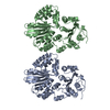
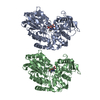
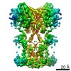
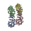
PROTEIN IS A CHIMERA COMPRISING MBP (UNP RESIDUES 27-395) AND THE C-TERMINAL DOMAIN (UNP RESIDUES ...PROTEIN IS A CHIMERA COMPRISING MBP (UNP RESIDUES 27-395) AND THE C-TERMINAL DOMAIN (UNP RESIDUES 263-294) OF SURVIVAL MOTOR NEURON PROTEIN.

-
Experimental details

-
Experiment

ExperimentMethod: X-RAY DIFFRACTION / Number of used crystals: 1

-
Sample preparation

CrystalDensity Matthews: 2.59 Å3/Da / Density % sol: 52.45 %
Crystal growTemperature: 294.15 K / Method: vapor diffusion, hanging drop / pH: 8
Details: 0.1 M Tris-HCl, pH 8.0, 14% w/v PEG3350, 10 mM calcium chloride, 0.1 M potassium chloride, 0.1 M ammonium sulfate, 18% ethylene glycol, VAPOR DIFFUSION, HANGING DROP, temperature 294.15K

-
Data collection

DiffractionMean temperature: 77 K
Diffraction sourceSource: SYNCHROTRON / Site: APS / Beamline: 24-ID-C / Wavelength: 0.9792 Å
DetectorType: ADSC QUANTUM 315 / Detector: CCD / Date: Feb 11, 2010
RadiationMonochromator: double crystal Si(111) / Protocol: SINGLE WAVELENGTH / Monochromatic (M) / Laue (L): M / Scattering type: x-ray
Radiation wavelengthWavelength: 0.9792 Å / Relative weight: 1
ReflectionResolution: 1.903→26.734 Å / Num. all: 35715 / Num. obs: 35714 / % possible obs: 99.4 % / Redundancy: 5.3 % / Biso Wilson estimate: 25.075 Å2 / Rmerge(I) obs: 0.126 / Net I/σ(I): 19.7
Reflection shellHighest resolution: 1.903 Å / Redundancy: 4.7 % / Rmerge(I) obs: 0.56 / Mean I/σ(I) obs: 2.4 / % possible all: 98.8

-
Processing

Software
NameVersionClassification
HKL-2000data collection
AMoREphasing
REFMAC5.5.0072refinement
HKL-2000data reduction
HKL-2000data scaling
RefinementMethod to determine structure: MOLECULAR REPLACEMENT
Starting model: PDB ENTRY 1OMP
Resolution: 1.903→26.734 Å / Cor.coef. Fo:Fc: 0.943 / Cor.coef. Fo:Fc free: 0.918 / SU B: 3.664 / SU ML: 0.11 / Cross valid method: THROUGHOUT / ESU R: 0.171 / ESU R Free: 0.159 / Stereochemistry target values: MAXIMUM LIKELIHOOD
Details: HYDROGENS HAVE BEEN ADDED IN THE RIDING POSITIONS U VALUES : REFINED INDIVIDUALLY
RfactorNum. reflection% reflectionSelection details
Rfree0.24709 -5 %RANDOM
Rwork0.20304 ---
all0.20533 35715 --
obs0.20533 35714 --
Solvent computationIon probe radii: 0.8 Å / Shrinkage radii: 0.8 Å / VDW probe radii: 1.4 Å / Solvent model: MASK
Displacement parametersBiso mean: 25.075 Å2
Baniso -1Baniso -2Baniso -3
1-1.16 Å20 Å21.63 Å2
2---1.75 Å20 Å2
3---1.73 Å2
Refinement stepCycle: LAST / Resolution: 1.903→26.734 Å
ProteinNucleic acidLigandSolventTotal
Num. atoms3033 0 0 160 3193
Refine LS restraints
Refine-IDTypeDev idealDev ideal targetNumber
X-RAY DIFFRACTIONr_bond_refined_d0.0240.0223136
X-RAY DIFFRACTIONr_angle_refined_deg1.9821.964258
X-RAY DIFFRACTIONr_dihedral_angle_1_deg6.5715393
X-RAY DIFFRACTIONr_dihedral_angle_2_deg37.30325.441136
X-RAY DIFFRACTIONr_dihedral_angle_3_deg17.86315530
X-RAY DIFFRACTIONr_dihedral_angle_4_deg16.45157
X-RAY DIFFRACTIONr_chiral_restr0.1590.2455
X-RAY DIFFRACTIONr_gen_planes_refined0.010.0212393
X-RAY DIFFRACTIONr_mcbond_it1.2571.51946
X-RAY DIFFRACTIONr_mcangle_it2.07323122
X-RAY DIFFRACTIONr_scbond_it3.38831190
X-RAY DIFFRACTIONr_scangle_it5.1054.51136
LS refinement shellResolution: 1.903→1.952 Å / Total num. of bins used: 20
RfactorNum. reflection% reflection
Rfree0.322 116 -
Rwork0.248 2175 -
obs--86.65 %

+
About Yorodumi

-
News

-
Feb 9, 2022. New format data for meta-information of EMDB entries

New format data for meta-information of EMDB entries

  • Version 3 of the EMDB header file is now the official format.
  • The previous official version 1.9 will be removed from the archive.

Related info.:EMDB header

External links:wwPDB to switch to version 3 of the EMDB data model

-
Aug 12, 2020. Covid-19 info

Covid-19 info

URL: https://pdbj.org/emnavi/covid19.php

New page: Covid-19 featured information page in EM Navigator.

Related info.:Covid-19 info / Mar 5, 2020. Novel coronavirus structure data

+
Mar 5, 2020. Novel coronavirus structure data

Novel coronavirus structure data

Related info.:Yorodumi Speices / Aug 12, 2020. Covid-19 info

External links:COVID-19 featured content - PDBj / Molecule of the Month (242):Coronavirus Proteases

+
Jan 31, 2019. EMDB accession codes are about to change! (news from PDBe EMDB page)

EMDB accession codes are about to change! (news from PDBe EMDB page)

  • The allocation of 4 digits for EMDB accession codes will soon come to an end. Whilst these codes will remain in use, new EMDB accession codes will include an additional digit and will expand incrementally as the available range of codes is exhausted. The current 4-digit format prefixed with “EMD-” (i.e. EMD-XXXX) will advance to a 5-digit format (i.e. EMD-XXXXX), and so on. It is currently estimated that the 4-digit codes will be depleted around Spring 2019, at which point the 5-digit format will come into force.
  • The EM Navigator/Yorodumi systems omit the EMD- prefix.

Related info.:Q: What is EMD? / ID/Accession-code notation in Yorodumi/EM Navigator

External links:EMDB Accession Codes are Changing Soon! / Contact to PDBj

+
Jul 12, 2017. Major update of PDB

Major update of PDB

  • wwPDB released updated PDB data conforming to the new PDBx/mmCIF dictionary.
  • This is a major update changing the version number from 4 to 5, and with Remediation, in which all the entries are updated.
  • In this update, many items about electron microscopy experimental information are reorganized (e.g. em_software).
  • Now, EM Navigator and Yorodumi are based on the updated data.

External links:wwPDB Remediation / Enriched Model Files Conforming to OneDep Data Standards Now Available in the PDB FTP Archive

-
Yorodumi

Thousand views of thousand structures

  • Yorodumi is a browser for structure data from EMDB, PDB, SASBDB, etc.
  • This page is also the successor to EM Navigator detail page, and also detail information page/front-end page for Omokage search.
  • The word "yorodu" (or yorozu) is an old Japanese word meaning "ten thousand". "mi" (miru) is to see.

Related info.:EMDB / PDB / SASBDB / Comparison of 3 databanks / Yorodumi Search / Aug 31, 2016. New EM Navigator & Yorodumi / Yorodumi Papers / Jmol/JSmol / Function and homology information / Changes in new EM Navigator and Yorodumi

Read more