Mass: 18.015 Da / Num. of mol.: 45 / Source method: isolated from a natural source / Formula: H2O
-
Experimental details
-
Experiment
Experiment
Method: X-RAY DIFFRACTION / Number of used crystals: 1
-
Sample preparation
Crystal
Density Matthews: 2.35 Å3/Da / Density % sol: 47.61 %
Crystal grow
Temperature: 291 K / Method: vapor diffusion, sitting drop / pH: 7.3 Details: 0.2 M HEPES, 7% MPEG5000, pH 7.3, VAPOR DIFFUSION, SITTING DROP, temperature 291K
-
Data collection
Diffraction
Mean temperature: 100 K
Diffraction source
Source: SYNCHROTRON / Type: SSRL / Wavelength: 0.95666 Å
Detector
Type: MAR / Detector: CCD / Date: Nov 16, 2008 Details: flat collimating Rh coated mirror, toroidal focusing mirror
Radiation
Monochromator: double crystal Si(111) / Protocol: SINGLE WAVELENGTH / Monochromatic (M) / Laue (L): M / Scattering type: x-ray
Radiation wavelength
Wavelength: 0.95666 Å / Relative weight: 1
Reflection
Redundancy: 9 % / Av σ(I) over netI: 32.5 / Number: 144772 / Rmerge(I) obs: 0.073 / Χ2: 1.26 / D res high: 2.6 Å / D res low: 50 Å / Num. obs: 16010 / % possible obs: 99.8
Diffraction reflection shell
Highest resolution (Å)
Lowest resolution (Å)
% possible obs (%)
ID
Rmerge(I) obs
Chi squared
Redundancy
5.6
50
98
1
0.036
1.197
8.5
4.45
5.6
100
1
0.046
1.144
9.3
3.88
4.45
99.9
1
0.061
1.113
9.1
3.53
3.88
100
1
0.087
1.33
8.9
3.28
3.53
100
1
0.12
1.372
8.9
3.08
3.28
100
1
0.147
1.136
9.3
2.93
3.08
100
1
0.226
1.002
9.3
2.8
2.93
100
1
0.349
1.043
9.3
2.69
2.8
100
1
0.543
1.196
9.2
2.6
2.69
100
1
0.788
2.172
8.7
Reflection
Resolution: 2.6→50 Å / Num. obs: 16010 / % possible obs: 99.8 % / Redundancy: 9 % / Rmerge(I) obs: 0.073 / Χ2: 1.262 / Net I/σ(I): 12.6
Reflection shell
Resolution (Å)
Redundancy (%)
Rmerge(I) obs
Num. unique all
Χ2
Diffraction-ID
% possible all
2.6-2.69
8.7
0.788
1552
2.172
1
100
2.69-2.8
9.2
0.543
1567
1.196
1
100
2.8-2.93
9.3
0.349
1567
1.043
1
100
2.93-3.08
9.3
0.226
1564
1.002
1
100
3.08-3.28
9.3
0.147
1577
1.136
1
100
3.28-3.53
8.9
0.12
1583
1.372
1
100
3.53-3.88
8.9
0.087
1610
1.33
1
100
3.88-4.45
9.1
0.061
1597
1.113
1
99.9
4.45-5.6
9.3
0.046
1644
1.144
1
100
5.6-50
8.5
0.036
1749
1.197
1
98
-
Phasing
Phasing
Method: molecular replacement
Phasing MR
Model details: Phaser MODE: MR_AUTO
Highest resolution
Lowest resolution
Rotation
2.69 Å
33.62 Å
Translation
2.69 Å
33.62 Å
-
Processing
Software
Name
Version
Classification
NB
DENZO
datareduction
SCALEPACK
datascaling
PHASER
2.3.0
phasing
REFMAC
refinement
PDB_EXTRACT
3.11
dataextraction
HKL-2000
datacollection
Refinement
Method to determine structure: MOLECULAR REPLACEMENT / Resolution: 2.75→27.5 Å / Cor.coef. Fo:Fc: 0.929 / Cor.coef. Fo:Fc free: 0.878 / WRfactor Rfree: 0.3293 / WRfactor Rwork: 0.2584 / Occupancy max: 1 / Occupancy min: 0.5 / FOM work R set: 0.7798 / SU B: 32.116 / SU ML: 0.321 / SU R Cruickshank DPI: 0.3743 / SU Rfree: 0.47 / Cross valid method: THROUGHOUT / σ(F): 0 / ESU R Free: 0.428 / Stereochemistry target values: MAXIMUM LIKELIHOOD / Details: HYDROGENS HAVE BEEN ADDED IN THE RIDING POSITIONS
Rfactor
Num. reflection
% reflection
Selection details
Rfree
0.2964
667
5 %
RANDOM
Rwork
0.2256
-
-
-
obs
0.229
13456
99.15 %
-
Solvent computation
Ion probe radii: 0.8 Å / Shrinkage radii: 0.8 Å / VDW probe radii: 1.2 Å / Solvent model: BABINET MODEL WITH MASK
In the structure databanks used in Yorodumi, some data are registered as the other names, "COVID-19 virus" and "2019-nCoV". Here are the details of the virus and the list of structure data.
Jan 31, 2019. EMDB accession codes are about to change! (news from PDBe EMDB page)
EMDB accession codes are about to change! (news from PDBe EMDB page)
The allocation of 4 digits for EMDB accession codes will soon come to an end. Whilst these codes will remain in use, new EMDB accession codes will include an additional digit and will expand incrementally as the available range of codes is exhausted. The current 4-digit format prefixed with “EMD-” (i.e. EMD-XXXX) will advance to a 5-digit format (i.e. EMD-XXXXX), and so on. It is currently estimated that the 4-digit codes will be depleted around Spring 2019, at which point the 5-digit format will come into force.
The EM Navigator/Yorodumi systems omit the EMD- prefix.
Related info.:Q: What is EMD? / ID/Accession-code notation in Yorodumi/EM Navigator
Yorodumi is a browser for structure data from EMDB, PDB, SASBDB, etc.
This page is also the successor to EM Navigator detail page, and also detail information page/front-end page for Omokage search.
The word "yorodu" (or yorozu) is an old Japanese word meaning "ten thousand". "mi" (miru) is to see.
Related info.:EMDB / PDB / SASBDB / Comparison of 3 databanks / Yorodumi Search / Aug 31, 2016. New EM Navigator & Yorodumi / Yorodumi Papers / Jmol/JSmol / Function and homology information / Changes in new EM Navigator and Yorodumi
 Movie
Movie Controller


 Open data
 Basic information
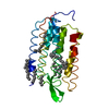
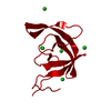
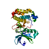
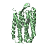
 Components
 Keywords
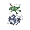
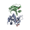
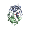
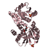
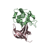
 beta barrel / OB-fold / protein-protein complex / novel scaffold / muraminidase /
  Function and homology information


 Authors
 Citation
 Structure visualization
 Downloads & links
 Other downloads


 PDBj
PDBj


 Assembly
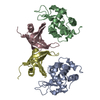


 Mass: 18.015 Da / Num. of mol.: 45 / Source method: isolated from a natural source / Formula: H2O
 Sample preparation
 / Wavelength: 0.95666 Å
 Processing