 Movie
Movie Controller

[English] 日本語
 Yorodumi
- PDB-4gcd: Room temperature X-ray diffraction study of a 6-fold molar excess... -
+ Open data

- Basic information
| Entry | Database: PDB / ID: 4gcd | ||||||
|---|---|---|---|---|---|---|---|
| Title | Room temperature X-ray diffraction study of a 6-fold molar excess of a cisplatin/carboplatin mixture binding to HEWL, Dataset 2 | ||||||
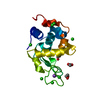
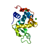
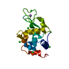
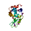
 Components Components | Lysozyme C | ||||||
 Keywords Keywords |  HYDROLASE / cisplatin / carboplatin / histidine / relative toxicity / binding occupancy / X-ray radiation damage / radiation therapy / temperature variation and structure HYDROLASE / cisplatin / carboplatin / histidine / relative toxicity / binding occupancy / X-ray radiation damage / radiation therapy / temperature variation and structure | ||||||
| Function / homology |  Function and homology informationAntimicrobial peptides / Neutrophil degranulation / beta-N-acetylglucosaminidase activity / cell wall macromolecule catabolic process / lysozyme / lysozyme activity / killing of cells of another organism / defense response to Gram-negative bacterium / defense response to Gram-positive bacterium / defense response to bacterium ...Antimicrobial peptides / Neutrophil degranulation / beta-N-acetylglucosaminidase activity / cell wall macromolecule catabolic process / lysozyme / lysozyme activity / killing of cells of another organism / defense response to Gram-negative bacterium / defense response to Gram-positive bacterium / defense response to bacterium / endoplasmic reticulum / extracellular space / identical protein binding / cytoplasm Function and homology informationAntimicrobial peptides / Neutrophil degranulation / beta-N-acetylglucosaminidase activity / cell wall macromolecule catabolic process / lysozyme / lysozyme activity / killing of cells of another organism / defense response to Gram-negative bacterium / defense response to Gram-positive bacterium / defense response to bacterium ...Antimicrobial peptides / Neutrophil degranulation / beta-N-acetylglucosaminidase activity / cell wall macromolecule catabolic process / lysozyme / lysozyme activity / killing of cells of another organism / defense response to Gram-negative bacterium / defense response to Gram-positive bacterium / defense response to bacterium / endoplasmic reticulum / extracellular space / identical protein binding / cytoplasmSimilarity search - Function | ||||||
| Biological species |  Gallus gallus (chicken) Gallus gallus (chicken) | ||||||
| Method | X-RAY DIFFRACTION / MOLECULAR REPLACEMENT / Resolution: 2.8 Å | ||||||
 Authors Authors | Helliwell, J.R. / Tanley, S.W.M. | ||||||
 Citation Citation | Journal: Acta Crystallogr.,Sect.D / Year: 2013 Title: The crystal structure analysis of the relative binding of cisplatin and carboplatin in a mixture with histidine in a protein studied at 100 and 300 K with repeated X-ray irradiation. Authors: Helliwell, J.R. / Tanley, S.W. | ||||||
| History |
|
- Structure visualization
| Structure viewer | Molecule: MolmilJmol/JSmol |
|---|
- Downloads & links
-Download
| PDBx/mmCIF format | 4gcd.cif.gz | 63.9 KB | Display | PDBx/mmCIF format |
|---|---|---|---|---|
| PDB format | pdb4gcd.ent.gz | 47 KB | Display | PDB format |
| PDBx/mmJSON format | 4gcd.json.gz | Tree view | PDBx/mmJSON format | |
| Others |  Other downloads Other downloads |
-Validation report
| Arichive directory | https://data.pdbj.org/pub/pdb/validation_reports/gc/4gcdftp://data.pdbj.org/pub/pdb/validation_reports/gc/4gcd | HTTPS FTP |
|---|
-Related structure data
| Related structure data |  4gcbC  4gccC  4gceC  4gcfC  2w1yS C: citing same article ( S: Starting model for refinement |
|---|---|
| Similar structure data |


-Links
 PDBj
PDBj


- Assembly
| Deposited unit | 
| ||||||||
|---|---|---|---|---|---|---|---|---|---|
| 1 |
| ||||||||
| Unit cell |
|
-Components
| #1: Protein | Mass: 14331.160 Da / Num. of mol.: 1 / Fragment: UNP residues 19-147 / Source method: isolated from a natural source / Source: (natural) Gallus gallus (chicken) / Tissue: egg white / References: UniProt: P00698, lysozyme | ||||
|---|---|---|---|---|---|
| #2: Chemical | Dimethyl sulfoxide  Mass: 78.133 Da / Num. of mol.: 3 / Source method: obtained synthetically / Formula: C2H6OS / Comment: DMSO, precipitant*YM Mass: 78.133 Da / Num. of mol.: 3 / Source method: obtained synthetically / Formula: C2H6OS / Comment: DMSO, precipitant*YM#3: Chemical | Cisplatin  Mass: 300.045 Da / Num. of mol.: 2 / Source method: obtained synthetically / Formula: Cl2H6N2Pt / Comment: medication, chemotherapy*YM Mass: 300.045 Da / Num. of mol.: 2 / Source method: obtained synthetically / Formula: Cl2H6N2Pt / Comment: medication, chemotherapy*YM#4: Water | ChemComp-HOH / | Water Mass: 18.015 Da / Num. of mol.: 6 / Source method: isolated from a natural source / Formula: H2O Mass: 18.015 Da / Num. of mol.: 6 / Source method: isolated from a natural source / Formula: H2O |
-Experimental details
-Experiment
| Experiment | Method: X-RAY DIFFRACTION / Number of used crystals: 1 |
|---|
- Sample preparation
| Crystal | Density Matthews: 2.09 Å3/Da / Density % sol: 41.22 % |
|---|---|
| Crystal grow | Temperature: 277 K / pH: 4.7 Details: 49 mg HEWL (3.2 mM), 3 mg cisplatin (10 mM), 3.7 mg carboplatin (10 mM) in 0.04 M sodium acetate buffer (462.5 uL) + 10% sodium chloride precipitant (462.5 uL) + 1 mM DMSO (75 uL), pH 4.7, ...Details: 49 mg HEWL (3.2 mM), 3 mg cisplatin (10 mM), 3.7 mg carboplatin (10 mM) in 0.04 M sodium acetate buffer (462.5 uL) + 10% sodium chloride precipitant (462.5 uL) + 1 mM DMSO (75 uL), pH 4.7, BATCH, temperature 277K |
-Data collection
| Diffraction | Mean temperature: 300 K |
|---|---|
| Diffraction source | Source: ROTATING ANODE / Type: BRUKER AXS MICROSTAR-H / Wavelength: 1.5418 Å |
| Detector | Type: APEX II CCD / Detector: CCD / Date: May 25, 2012 |
| Radiation | Monochromator: confocal mirror optics / Protocol: SINGLE WAVELENGTH / Monochromatic (M) / Laue (L): M / Scattering type: x-ray |
| Radiation wavelength | Wavelength: 1.5418 Å / Relative weight: 1 |
| Reflection | Resolution: 2.8→35.74 Å / Num. all: 3140 / Num. obs: 3141 / % possible obs: 100 % / Observed criterion σ(F): 4 / Observed criterion σ(I): 2 / Redundancy: 23.6 % / Rmerge(I) obs: 0.289 / Net I/σ(I): 10.8 |
| Reflection shell | Resolution: 2.8→2.87 Å / Redundancy: 25.7 % / Rmerge(I) obs: 0.575 / Mean I/σ(I) obs: 4.5 / % possible all: 100 |
- Processing
| Software |
| ||||||||||||||||||||||||||||||||||||||||||||||||||||||||||||
|---|---|---|---|---|---|---|---|---|---|---|---|---|---|---|---|---|---|---|---|---|---|---|---|---|---|---|---|---|---|---|---|---|---|---|---|---|---|---|---|---|---|---|---|---|---|---|---|---|---|---|---|---|---|---|---|---|---|---|---|---|---|
| Refinement | Method to determine structure: MOLECULAR REPLACEMENT Starting model: PDB ENTRY 2W1Y Resolution: 2.8→35.74 Å / Cor.coef. Fo:Fc: 0.946 / Cor.coef. Fo:Fc free: 0.913 / SU B: 34.073 / SU ML: 0.314 / Cross valid method: THROUGHOUT / σ(F): 4 / σ(I): 2 / ESU R Free: 0.39 / Stereochemistry target values: MAXIMUM LIKELIHOOD Details: HYDROGENS HAVE BEEN ADDED IN THE RIDING POSITIONS U VALUES : REFINED INDIVIDUALLY
| ||||||||||||||||||||||||||||||||||||||||||||||||||||||||||||
| Solvent computation | Ion probe radii: 0.8 Å / Shrinkage radii: 0.8 Å / VDW probe radii: 1.2 Å / Solvent model: BABINET MODEL WITH MASK | ||||||||||||||||||||||||||||||||||||||||||||||||||||||||||||
| Displacement parameters | Biso mean: 34.654 Å2
| ||||||||||||||||||||||||||||||||||||||||||||||||||||||||||||
| Refinement step | Cycle: LAST / Resolution: 2.8→35.74 Å
| ||||||||||||||||||||||||||||||||||||||||||||||||||||||||||||
| Refine LS restraints |
| ||||||||||||||||||||||||||||||||||||||||||||||||||||||||||||
| LS refinement shell | Resolution: 2.8→2.873 Å / Total num. of bins used: 20
| ||||||||||||||||||||||||||||||||||||||||||||||||||||||||||||
| Refinement TLS params. | Method: refined / Origin x: -20.7943 Å / Origin y: -0.5023 Å / Origin z: -9.3736 Å
|
