 Movie
Movie Controller


+ Open data

- Basic information
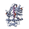
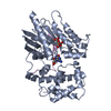
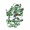
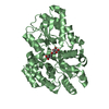
| Entry | Database: PDB / ID: 4g5r | ||||||
|---|---|---|---|---|---|---|---|
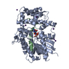
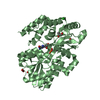
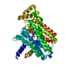
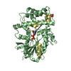
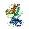
| Title | Structure of LGN GL4/Galphai3 complex | ||||||
 Components Components |
| ||||||
 Keywords Keywords | CELL CYCLE/SIGNALING PROTEIN / Galphai / GoLoco / Galpha signaling /  asymmetric cell division / CELL CYCLE-SIGNALING PROTEIN complex asymmetric cell division / CELL CYCLE-SIGNALING PROTEIN complex | ||||||
| Function / homology |  Function and homology information Function and homology informationRan protein signal transduction / lateral cell cortex / cell cortex region / maintenance of centrosome location / positive regulation of spindle assembly / G alpha (i) signalling events / negative regulation of adenylate cyclase activity / GTP metabolic process / GDP-dissociation inhibitor activity / dopamine receptor signaling pathway ...Ran protein signal transduction / lateral cell cortex / cell cortex region / maintenance of centrosome location / positive regulation of spindle assembly / G alpha (i) signalling events / negative regulation of adenylate cyclase activity / GTP metabolic process / GDP-dissociation inhibitor activity / dopamine receptor signaling pathway / dynein complex binding / mitotic spindle pole / establishment of mitotic spindle orientation / positive regulation of macroautophagy / Adenylate cyclase inhibitory pathway / positive regulation of protein localization to cell cortex / G-protein alpha-subunit binding / lateral plasma membrane / regulation of mitotic spindle organization / adenylate cyclase-inhibiting G protein-coupled receptor signaling pathway / mitotic spindle organization / G protein-coupled receptor binding / G-protein beta/gamma-subunit complex binding / adenylate cyclase-modulating G protein-coupled receptor signaling pathway / ADP signalling through P2Y purinoceptor 12 / G alpha (z) signalling events / ADORA2B mediated anti-inflammatory cytokines production / GPER1 signaling / GDP binding / heterotrimeric G-protein complex / : / cell cortex / midbody / G alpha (i) signalling events / G alpha (s) signalling events / Extra-nuclear estrogen signaling / cell cycle / cell division / lysosomal membrane / protein domain specific binding / nucleotide binding / GTPase activity / centrosome / GTP binding / nucleolus / Golgi apparatus / protein-containing complex / extracellular exosome / nucleoplasm / membrane / identical protein binding / metal ion binding / plasma membrane / cytosol / cytoplasmSimilarity search - Function | ||||||
| Biological species |  Homo sapiens (human) Homo sapiens (human) Mus musculus (house mouse) Mus musculus (house mouse) | ||||||
| Method | X-RAY DIFFRACTION / SYNCHROTRON / Resolution: 3.481 Å | ||||||
 Authors Authors | Jia, M. / Li, J. / Zhu, J. / Wen, W. / Zhang, M. / Wang, W. | ||||||
 Citation Citation | Journal: To be Published Title: Crystal Structures of the scaffolding protein LGN reveal the general mechanism by which GoLoco binding motifs inhibit the release of GDP from Galphai subunits in G-coupled heterotrimeric proteins Authors: Jia, M. / Li, J. / Zhu, J. / Wen, W. / Zhang, M. / Wang, W. | ||||||
| History |
|
- Structure visualization
| Structure viewer | Molecule: MolmilJmol/JSmol |
|---|
- Downloads & links
-Download
| PDBx/mmCIF format | 4g5r.cif.gz | 275.3 KB | Display | PDBx/mmCIF format |
|---|---|---|---|---|
| PDB format | pdb4g5r.ent.gz | 220.3 KB | Display | PDB format |
| PDBx/mmJSON format | 4g5r.json.gz | Tree view | PDBx/mmJSON format | |
| Others |  Other downloads Other downloads |
-Validation report
| Arichive directory | https://data.pdbj.org/pub/pdb/validation_reports/g5/4g5rftp://data.pdbj.org/pub/pdb/validation_reports/g5/4g5r | HTTPS FTP |
|---|
-Related structure data


-Links
 PDBj
PDBj


- Assembly
| Deposited unit | 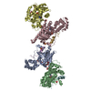
| ||||||||
|---|---|---|---|---|---|---|---|---|---|
| 1 | 
| ||||||||
| 2 | 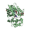
| ||||||||
| 3 | 
| ||||||||
| 4 | 
| ||||||||
| Unit cell |
|
-Components
| #1: Protein | Mass: 37903.023 Da / Num. of mol.: 4 / Fragment: UNP RESIDUES 25-354 Source method: isolated from a genetically manipulated source Source: (gene. exp.) Homo sapiens (human) / Gene: GNAI3 / Production host:  Escherichia coli (E. coli) / References: UniProt: P08754 Escherichia coli (E. coli) / References: UniProt: P08754#2: Protein/peptide | Mass: 3099.544 Da / Num. of mol.: 4 / Fragment: GoLoco 4 (UNP RESIDUES 628-653) / Source method: obtained synthetically / Details: This sequence occurs naturally in mouse. / Source: (synth.) Mus musculus (house mouse) / References: UniProt: Q8VDU0#3: Chemical | ChemComp-GDP / Guanosine diphosphate  Type: RNA linking / Mass: 443.201 Da / Num. of mol.: 4 / Source method: obtained synthetically / Formula: C10H15N5O11P2 / Comment: GDP, energy-carrying molecule*YM Type: RNA linking / Mass: 443.201 Da / Num. of mol.: 4 / Source method: obtained synthetically / Formula: C10H15N5O11P2 / Comment: GDP, energy-carrying molecule*YM#4: Chemical | Sulfate  Mass: 96.063 Da / Num. of mol.: 3 / Source method: obtained synthetically / Formula: SO4 Mass: 96.063 Da / Num. of mol.: 3 / Source method: obtained synthetically / Formula: SO4#5: Chemical | ChemComp-CIT / Citric acid  Mass: 192.124 Da / Num. of mol.: 4 / Source method: obtained synthetically / Formula: C6H8O7 Mass: 192.124 Da / Num. of mol.: 4 / Source method: obtained synthetically / Formula: C6H8O7 |
|---|
-Experimental details
-Experiment
| Experiment | Method: X-RAY DIFFRACTION / Number of used crystals: 1 |
|---|
- Sample preparation
| Crystal | Density Matthews: 4.58 Å3/Da / Density % sol: 73.16 % |
|---|---|
| Crystal grow | Temperature: 291 K / Method: vapor diffusion, hanging drop / pH: 5.6 Details: 0.5M ammonium sulfate, 1.0M lithium sulfate monohydrate, 0.1M sodium citrate tribasic dihydrate, pH 5.6, VAPOR DIFFUSION, HANGING DROP, temperature 291K |
-Data collection
| Diffraction | Mean temperature: 100 K |
|---|---|
| Diffraction source | Source: SYNCHROTRON / Site: SSRF  / Beamline: BL17U / Wavelength: 0.9793 Å / Beamline: BL17U / Wavelength: 0.9793 Å |
| Detector | Type: ADSC QUANTUM 315r / Detector: CCD / Date: Nov 6, 2010 |
| Radiation | Monochromator: Si(111) / Protocol: SINGLE WAVELENGTH / Monochromatic (M) / Laue (L): M / Scattering type: x-ray |
| Radiation wavelength | Wavelength: 0.9793 Å / Relative weight: 1 |
| Reflection | Resolution: 3.48→50 Å / Num. all: 39388 / Num. obs: 39267 / Observed criterion σ(F): 2 / Observed criterion σ(I): 2 / Redundancy: 4.4 % / Biso Wilson estimate: 95.39 Å2 / Rmerge(I) obs: 0.11 / Net I/σ(I): 14.7 |
| Reflection shell | Resolution: 3.5→3.56 Å / Redundancy: 4.5 % / Rmerge(I) obs: 0.757 / Mean I/σ(I) obs: 1.8 / Num. unique all: 1955 |
- Processing
| Software |
| |||||||||||||||||||||||||||||||||||||||||||||||||||||||||||||||||||||||||||||||||||||||||||||||||||||||||
|---|---|---|---|---|---|---|---|---|---|---|---|---|---|---|---|---|---|---|---|---|---|---|---|---|---|---|---|---|---|---|---|---|---|---|---|---|---|---|---|---|---|---|---|---|---|---|---|---|---|---|---|---|---|---|---|---|---|---|---|---|---|---|---|---|---|---|---|---|---|---|---|---|---|---|---|---|---|---|---|---|---|---|---|---|---|---|---|---|---|---|---|---|---|---|---|---|---|---|---|---|---|---|---|---|---|---|
| Refinement | Resolution: 3.481→48.044 Å / Occupancy max: 1 / Occupancy min: 1 / FOM work R set: 0.8099 / SU ML: 0.4 / σ(F): 1.35 / Phase error: 25.26 / Stereochemistry target values: ML
| |||||||||||||||||||||||||||||||||||||||||||||||||||||||||||||||||||||||||||||||||||||||||||||||||||||||||
| Solvent computation | Shrinkage radii: 0.9 Å / VDW probe radii: 1.11 Å / Solvent model: FLAT BULK SOLVENT MODEL | |||||||||||||||||||||||||||||||||||||||||||||||||||||||||||||||||||||||||||||||||||||||||||||||||||||||||
| Displacement parameters | Biso max: 199.31 Å2 / Biso mean: 103.2263 Å2 / Biso min: 37.1 Å2 | |||||||||||||||||||||||||||||||||||||||||||||||||||||||||||||||||||||||||||||||||||||||||||||||||||||||||
| Refinement step | Cycle: LAST / Resolution: 3.481→48.044 Å
| |||||||||||||||||||||||||||||||||||||||||||||||||||||||||||||||||||||||||||||||||||||||||||||||||||||||||
| Refine LS restraints |
| |||||||||||||||||||||||||||||||||||||||||||||||||||||||||||||||||||||||||||||||||||||||||||||||||||||||||
| LS refinement shell | Refine-ID: X-RAY DIFFRACTION / Total num. of bins used: 14
|
