 Movie
Movie Controller


+ Open data

- Basic information
| Entry | Database: PDB / ID: 4f2x | ||||||
|---|---|---|---|---|---|---|---|
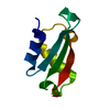
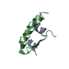
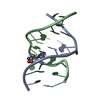
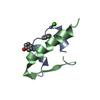
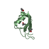
| Title | Structure of 3'-Fluoro Cyclohexenyl Nucleic Acid Heptamer | ||||||
 Components Components |
| ||||||
 Keywords Keywords |  DNA / Fluoro Cyclohexenyl Nucleic Acid / F-CeNA / Modified DNA / B-form DNA DNA / Fluoro Cyclohexenyl Nucleic Acid / F-CeNA / Modified DNA / B-form DNA | ||||||
| Function / homology | COBALT (III) ION / COBALT HEXAMMINE(III) / DNA Function and homology information Function and homology information | ||||||
| Method | X-RAY DIFFRACTION / SYNCHROTRON / MOLECULAR REPLACEMENT / Resolution: 1.57 Å | ||||||
 Authors Authors | Pallan, P.S. / Egli, M. | ||||||
 Citation Citation | Journal: J.Org.Chem. / Year: 2012 Title: Synthesis and Antisense Properties of Fluoro Cyclohexenyl Nucleic Acid (F-CeNA), a Nuclease Stable Mimic of 2'-Fluoro RNA. Authors: Seth, P.P. / Yu, J. / Jazayeri, A. / Pallan, P.S. / Allerson, C.R. / Ostergaard, M.E. / Liu, F. / Herdewijn, P. / Egli, M. / Swayze, E.E. | ||||||
| History |
|
- Structure visualization
| Structure viewer | Molecule: MolmilJmol/JSmol |
|---|
- Downloads & links
-Download
| PDBx/mmCIF format | 4f2x.cif.gz | 41.1 KB | Display | PDBx/mmCIF format |
|---|---|---|---|---|
| PDB format | pdb4f2x.ent.gz | 30.1 KB | Display | PDB format |
| PDBx/mmJSON format | 4f2x.json.gz | Tree view | PDBx/mmJSON format | |
| Others |  Other downloads Other downloads |
-Validation report
| Arichive directory | https://data.pdbj.org/pub/pdb/validation_reports/f2/4f2xftp://data.pdbj.org/pub/pdb/validation_reports/f2/4f2x | HTTPS FTP |
|---|
-Related structure data
| Related structure data | 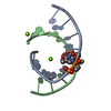 4f2yC  3fl6S C: citing same article ( S: Starting model for refinement |
|---|---|
| Similar structure data |


-Links
 PDBj
PDBj


- Assembly
| Deposited unit | 
| ||||||||
|---|---|---|---|---|---|---|---|---|---|
| 1 | 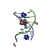
| ||||||||
| 2 | 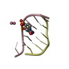
| ||||||||
| Unit cell |
|
-Components
| #1: DNA chain | Mass: 2182.451 Da / Num. of mol.: 2 / Source method: obtained synthetically #2: DNA chain | Mass: 2083.388 Da / Num. of mol.: 2 / Source method: obtained synthetically #3: Chemical |   Mass: 161.116 Da / Num. of mol.: 2 / Source method: obtained synthetically / Formula: CoH18N6 Mass: 161.116 Da / Num. of mol.: 2 / Source method: obtained synthetically / Formula: CoH18N6#4: Chemical | ChemComp-3CO / Cobalt  Mass: 58.933 Da / Num. of mol.: 6 / Source method: obtained synthetically / Formula: Co Mass: 58.933 Da / Num. of mol.: 6 / Source method: obtained synthetically / Formula: Co#5: Water | ChemComp-HOH / | Water Mass: 18.015 Da / Num. of mol.: 77 / Source method: isolated from a natural source / Formula: H2O Mass: 18.015 Da / Num. of mol.: 77 / Source method: isolated from a natural source / Formula: H2O |
|---|
-Experimental details
-Experiment
| Experiment | Method: X-RAY DIFFRACTION / Number of used crystals: 1 |
|---|
- Sample preparation
| Crystal | Density Matthews: 2.12 Å3/Da / Density % sol: 42.08 % |
|---|---|
| Crystal grow | Temperature: 291 K / Method: vapor diffusion, hanging drop / pH: 5.5 Details: 40 mM sodium cacodylate, 20 mM cobalt hexamine, 20 mM magnesium chloride, 10% v/v MPD, pH 5.5, VAPOR DIFFUSION, HANGING DROP, temperature 291K |
-Data collection
| Diffraction | Mean temperature: 100 K |
|---|---|
| Diffraction source | Source: SYNCHROTRON / Site: APS  / Beamline: 21-ID-F / Wavelength: 0.9787 Å / Beamline: 21-ID-F / Wavelength: 0.9787 Å |
| Detector | Type: MARMOSAIC 225 mm CCD / Detector: CCD / Date: Feb 7, 2012 |
| Radiation | Monochromator: diamond(111) / Protocol: SINGLE WAVELENGTH / Monochromatic (M) / Laue (L): M / Scattering type: x-ray |
| Radiation wavelength | Wavelength: 0.9787 Å / Relative weight: 1 |
| Reflection | Resolution: 1.57→40.08 Å / Num. all: 10628 / Num. obs: 10592 / % possible obs: 99.6 % / Observed criterion σ(I): 5 / Redundancy: 7.7 % / Rmerge(I) obs: 0.063 / Net I/σ(I): 38.6 |
| Reflection shell | Resolution: 1.57→1.63 Å / Redundancy: 7.5 % / Rmerge(I) obs: 0.59 / Mean I/σ(I) obs: 3.63 / % possible all: 99.3 |
- Processing
| Software |
| |||||||||||||||||||||||||||||||||||||||||||||||||||||||||||||||||||||||||||
|---|---|---|---|---|---|---|---|---|---|---|---|---|---|---|---|---|---|---|---|---|---|---|---|---|---|---|---|---|---|---|---|---|---|---|---|---|---|---|---|---|---|---|---|---|---|---|---|---|---|---|---|---|---|---|---|---|---|---|---|---|---|---|---|---|---|---|---|---|---|---|---|---|---|---|---|---|
| Refinement | Method to determine structure: MOLECULAR REPLACEMENT Starting model: PDB ENTRY 3FL6 Resolution: 1.57→40.08 Å / Cor.coef. Fo:Fc: 0.96 / Cor.coef. Fo:Fc free: 0.933 / SU B: 3.219 / SU ML: 0.071 / Cross valid method: THROUGHOUT / ESU R: 0.098 / ESU R Free: 0.104 / Stereochemistry target values: MAXIMUM LIKELIHOOD Details: HYDROGENS HAVE BEEN USED IF PRESENT IN THE INPUT U VALUES : RESIDUAL ONLY
| |||||||||||||||||||||||||||||||||||||||||||||||||||||||||||||||||||||||||||
| Solvent computation | Ion probe radii: 0.8 Å / Shrinkage radii: 0.8 Å / VDW probe radii: 1.2 Å / Solvent model: MASK | |||||||||||||||||||||||||||||||||||||||||||||||||||||||||||||||||||||||||||
| Displacement parameters | Biso mean: 25.805 Å2
| |||||||||||||||||||||||||||||||||||||||||||||||||||||||||||||||||||||||||||
| Refinement step | Cycle: LAST / Resolution: 1.57→40.08 Å
| |||||||||||||||||||||||||||||||||||||||||||||||||||||||||||||||||||||||||||
| Refine LS restraints |
| |||||||||||||||||||||||||||||||||||||||||||||||||||||||||||||||||||||||||||
| LS refinement shell | Resolution: 1.57→1.612 Å / Total num. of bins used: 20
| |||||||||||||||||||||||||||||||||||||||||||||||||||||||||||||||||||||||||||
| Refinement TLS params. | Method: refined / Refine-ID: X-RAY DIFFRACTION
| |||||||||||||||||||||||||||||||||||||||||||||||||||||||||||||||||||||||||||
| Refinement TLS group |
|
