 Movie
Movie Controller

[English] 日本語
 Yorodumi
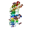
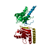
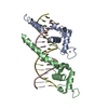
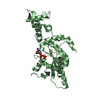
- PDB-4etx: Crystal Structure of PelD 158-CT from Pseudomonas aeruginosa PAO1 -
+ Open data

- Basic information
| Entry | Database: PDB / ID: 4etx | ||||||
|---|---|---|---|---|---|---|---|
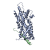
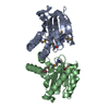
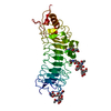
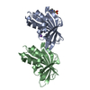
| Title | Crystal Structure of PelD 158-CT from Pseudomonas aeruginosa PAO1 | ||||||
 Components Components | PelD Pediatric end-stage liver disease Pediatric end-stage liver disease | ||||||
 Keywords Keywords | SIGNALING PROTEIN / c-di-GMP | ||||||
| Function / homology |  Function and homology information Function and homology informationextracellular polysaccharide biosynthetic process / cyclic-di-GMP binding / single-species biofilm formation / membraneSimilarity search - Function | ||||||
| Biological species |   Pseudomonas aeruginosa (bacteria) Pseudomonas aeruginosa (bacteria) | ||||||
| Method | X-RAY DIFFRACTION / SYNCHROTRON / SAD / Resolution: 2 Å | ||||||
 Authors Authors | Li, Z. / Chen, J. / Nair, S.K. | ||||||
 Citation Citation | Journal: J.Biol.Chem. / Year: 2012 Title: Structures of the PelD Cyclic Diguanylate Effector Involved in Pellicle Formation in Pseudomonas aeruginosa PAO1. Authors: Li, Z. / Chen, J.H. / Hao, Y. / Nair, S.K. | ||||||
| History |
|
- Structure visualization
| Structure viewer | Molecule: MolmilJmol/JSmol |
|---|
- Downloads & links
-Download
| PDBx/mmCIF format | 4etx.cif.gz | 131.1 KB | Display | PDBx/mmCIF format |
|---|---|---|---|---|
| PDB format | pdb4etx.ent.gz | 102.9 KB | Display | PDB format |
| PDBx/mmJSON format | 4etx.json.gz | Tree view | PDBx/mmJSON format | |
| Others |  Other downloads Other downloads |
-Validation report
| Arichive directory | https://data.pdbj.org/pub/pdb/validation_reports/et/4etxftp://data.pdbj.org/pub/pdb/validation_reports/et/4etx | HTTPS FTP |
|---|
-Related structure data


-Links
 PDBj
PDBj

- Assembly
| Deposited unit | 
| ||||||||
|---|---|---|---|---|---|---|---|---|---|
| 1 |
| ||||||||
| Unit cell |
|
-Components
| #1: Protein | Pediatric end-stage liver disease Mass: 33712.344 Da / Num. of mol.: 1 / Fragment: UNP residues 155-454 Source method: isolated from a genetically manipulated source Source: (gene. exp.) Pseudomonas aeruginosa (bacteria) / Strain: PAO1 / Gene: PA3061, pelD / Plasmid: pET28 / Production host: Escherichia coli (E. coli) / Strain (production host): BL21 Rosetta(DE3) / References: UniProt: Q9HZE7 |
|---|---|
| #2: Water | ChemComp-HOH / Water Mass: 18.015 Da / Num. of mol.: 86 / Source method: isolated from a natural source / Formula: H2O Mass: 18.015 Da / Num. of mol.: 86 / Source method: isolated from a natural source / Formula: H2O |
-Experimental details
-Experiment
| Experiment | Method: X-RAY DIFFRACTION / Number of used crystals: 1 |
|---|
- Sample preparation
| Crystal | Density Matthews: 2.12 Å3/Da / Density % sol: 41.87 % |
|---|---|
| Crystal grow | Temperature: 298 K / Method: hanging drop / pH: 8 Details: 100 mM Tris, pH 8, 200 mM MgCl2, 10% (v/v) PEG 8000, hanging drop, temperature 298K |
-Data collection
| Diffraction source | Source: SYNCHROTRON / Site: APS  / Beamline: 21-ID-F / Wavelength: 1 Å / Beamline: 21-ID-F / Wavelength: 1 Å | |||||||||||||||||||||||||||||||||||||||||||||||||||||||||||||||||||||||||||||||||||||||||||
|---|---|---|---|---|---|---|---|---|---|---|---|---|---|---|---|---|---|---|---|---|---|---|---|---|---|---|---|---|---|---|---|---|---|---|---|---|---|---|---|---|---|---|---|---|---|---|---|---|---|---|---|---|---|---|---|---|---|---|---|---|---|---|---|---|---|---|---|---|---|---|---|---|---|---|---|---|---|---|---|---|---|---|---|---|---|---|---|---|---|---|---|---|
| Detector | Type: MAR scanner 300 mm plate / Detector: IMAGE PLATE | |||||||||||||||||||||||||||||||||||||||||||||||||||||||||||||||||||||||||||||||||||||||||||
| Radiation | Protocol: SINGLE WAVELENGTH / Monochromatic (M) / Laue (L): M / Scattering type: x-ray | |||||||||||||||||||||||||||||||||||||||||||||||||||||||||||||||||||||||||||||||||||||||||||
| Radiation wavelength | Wavelength: 1 Å / Relative weight: 1 | |||||||||||||||||||||||||||||||||||||||||||||||||||||||||||||||||||||||||||||||||||||||||||
| Reflection | Highest resolution: 2 Å / Num. obs: 19307 / % possible obs: 99.8 % / Observed criterion σ(I): -3 / Biso Wilson estimate: 41.15 Å2 / Rmerge(I) obs: 0.047 / Net I/σ(I): 24.69 | |||||||||||||||||||||||||||||||||||||||||||||||||||||||||||||||||||||||||||||||||||||||||||
| Reflection shell | Diffraction-ID: 1
|
- Processing
| Software |
| |||||||||||||||||||||||||||||||||||||||||||||||||||||||||||||||||
|---|---|---|---|---|---|---|---|---|---|---|---|---|---|---|---|---|---|---|---|---|---|---|---|---|---|---|---|---|---|---|---|---|---|---|---|---|---|---|---|---|---|---|---|---|---|---|---|---|---|---|---|---|---|---|---|---|---|---|---|---|---|---|---|---|---|---|
| Refinement | Method to determine structure: SAD / Resolution: 2→25 Å / Cor.coef. Fo:Fc: 0.938 / Cor.coef. Fo:Fc free: 0.897 / Occupancy max: 1 / Occupancy min: 1 / SU B: 12.015 / SU ML: 0.155 / Cross valid method: THROUGHOUT / σ(F): 0 / ESU R: 0.254 / ESU R Free: 0.212 / Stereochemistry target values: MAXIMUM LIKELIHOOD Details: HYDROGENS HAVE BEEN ADDED IN THE RIDING POSITIONS U VALUES : RESIDUAL ONLY
| |||||||||||||||||||||||||||||||||||||||||||||||||||||||||||||||||
| Solvent computation | Ion probe radii: 0.8 Å / Shrinkage radii: 0.8 Å / VDW probe radii: 1.4 Å / Solvent model: BABINET MODEL WITH MASK | |||||||||||||||||||||||||||||||||||||||||||||||||||||||||||||||||
| Displacement parameters | Biso max: 84.25 Å2 / Biso mean: 42.27 Å2 / Biso min: 8.62 Å2
| |||||||||||||||||||||||||||||||||||||||||||||||||||||||||||||||||
| Refinement step | Cycle: LAST / Resolution: 2→25 Å
| |||||||||||||||||||||||||||||||||||||||||||||||||||||||||||||||||
| Refine LS restraints |
| |||||||||||||||||||||||||||||||||||||||||||||||||||||||||||||||||
| LS refinement shell | Resolution: 2→2.052 Å / Total num. of bins used: 20
| |||||||||||||||||||||||||||||||||||||||||||||||||||||||||||||||||
| Refinement TLS params. | Method: refined / Origin x: -4.2278 Å / Origin y: 1.5679 Å / Origin z: 15.0848 Å
|
