 Movie
Movie Controller


+ Open data

- Basic information
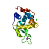
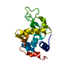
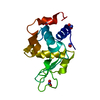
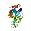
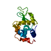
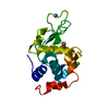
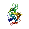
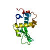
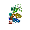
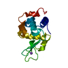
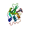
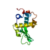
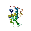
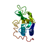
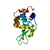
| Entry | Database: PDB / ID: 4etb | ||||||
|---|---|---|---|---|---|---|---|
| Title | lysozyme, room temperature, 200 kGy dose | ||||||
 Components Components | Lysozyme C | ||||||
 Keywords Keywords |  HYDROLASE / lysozyme HYDROLASE / lysozyme | ||||||
| Function / homology |  Function and homology informationAntimicrobial peptides / Neutrophil degranulation / beta-N-acetylglucosaminidase activity / cell wall macromolecule catabolic process / lysozyme / lysozyme activity / killing of cells of another organism / defense response to Gram-negative bacterium / defense response to Gram-positive bacterium / defense response to bacterium ...Antimicrobial peptides / Neutrophil degranulation / beta-N-acetylglucosaminidase activity / cell wall macromolecule catabolic process / lysozyme / lysozyme activity / killing of cells of another organism / defense response to Gram-negative bacterium / defense response to Gram-positive bacterium / defense response to bacterium / endoplasmic reticulum / extracellular space / identical protein binding / cytoplasm Function and homology informationAntimicrobial peptides / Neutrophil degranulation / beta-N-acetylglucosaminidase activity / cell wall macromolecule catabolic process / lysozyme / lysozyme activity / killing of cells of another organism / defense response to Gram-negative bacterium / defense response to Gram-positive bacterium / defense response to bacterium ...Antimicrobial peptides / Neutrophil degranulation / beta-N-acetylglucosaminidase activity / cell wall macromolecule catabolic process / lysozyme / lysozyme activity / killing of cells of another organism / defense response to Gram-negative bacterium / defense response to Gram-positive bacterium / defense response to bacterium / endoplasmic reticulum / extracellular space / identical protein binding / cytoplasmSimilarity search - Function | ||||||
| Biological species |  Gallus gallus (chicken) Gallus gallus (chicken) | ||||||
| Method | X-RAY DIFFRACTION / SYNCHROTRON / FOURIER SYNTHESIS / Resolution: 1.908 Å | ||||||
 Authors Authors | Boutet, S. / Lomb, L. / Williams, G. / Barends, T. / Aquila, A. / Doak, R.B. / Weierstall, U. / DePonte, D. / Steinbrener, J. / Shoeman, R. ...Boutet, S. / Lomb, L. / Williams, G. / Barends, T. / Aquila, A. / Doak, R.B. / Weierstall, U. / DePonte, D. / Steinbrener, J. / Shoeman, R. / Messerschmidt, M. / Barty, A. / White, T. / Kassemeyer, S. / Kirian, R. / Seibert, M. / Montanez, P. / Kenney, C. / Herbst, R. / Hart, P. / Pines, J. / Haller, G. / Gruner, S. / Philllip, H. / Tate, M. / Hromalik, M. / Koerner, L. / van Bakel, N. / Morse, J. / Ghonsalves, W. / Arnlund, D. / Bogan, M. / Calemann, C. / Fromme, R. / Hampton, C. / Hunter, M. / Johansson, L. / Katona, G. / Kupitz, C. / Liang, M. / Martin, A. / Nass, K. / Redecke, L. / Stellato, F. / Timneanu, N. / Wang, D. / Zatsepin, N. / Schafer, D. / Defever, K. / Neutze, R. / Fromme, P. / Spence, J. / Chapman, H. / Schlichting, I. | ||||||
 Citation Citation | Journal: Science / Year: 2012 Title: High-resolution protein structure determination by serial femtosecond crystallography. Authors: Boutet, S. / Lomb, L. / Williams, G.J. / Barends, T.R. / Aquila, A. / Doak, R.B. / Weierstall, U. / DePonte, D.P. / Steinbrener, J. / Shoeman, R.L. / Messerschmidt, M. / Barty, A. / White, T. ...Authors: Boutet, S. / Lomb, L. / Williams, G.J. / Barends, T.R. / Aquila, A. / Doak, R.B. / Weierstall, U. / DePonte, D.P. / Steinbrener, J. / Shoeman, R.L. / Messerschmidt, M. / Barty, A. / White, T.A. / Kassemeyer, S. / Kirian, R.A. / Seibert, M.M. / Montanez, P.A. / Kenney, C. / Herbst, R. / Hart, P. / Pines, J. / Haller, G. / Gruner, S.M. / Philipp, H.T. / Tate, M.W. / Hromalik, M. / Koerner, L.J. / van Bakel, N. / Morse, J. / Ghonsalves, W. / Arnlund, D. / Bogan, M.J. / Caleman, C. / Fromme, R. / Hampton, C.Y. / Hunter, M.S. / Johansson, L.C. / Katona, G. / Kupitz, C. / Liang, M. / Martin, A.V. / Nass, K. / Redecke, L. / Stellato, F. / Timneanu, N. / Wang, D. / Zatsepin, N.A. / Schafer, D. / Defever, J. / Neutze, R. / Fromme, P. / Spence, J.C. / Chapman, H.N. / Schlichting, I. | ||||||
| History |
|
- Structure visualization
| Structure viewer | Molecule: MolmilJmol/JSmol |
|---|
- Downloads & links
-Download
| PDBx/mmCIF format | 4etb.cif.gz | 35 KB | Display | PDBx/mmCIF format |
|---|---|---|---|---|
| PDB format | pdb4etb.ent.gz | 26.3 KB | Display | PDB format |
| PDBx/mmJSON format | 4etb.json.gz | Tree view | PDBx/mmJSON format | |
| Others |  Other downloads Other downloads |
-Validation report
| Arichive directory | https://data.pdbj.org/pub/pdb/validation_reports/et/4etbftp://data.pdbj.org/pub/pdb/validation_reports/et/4etb | HTTPS FTP |
|---|
-Related structure data
| Related structure data | 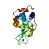 4et8C  4et9C 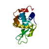 4etaC  4etcC  4etdC 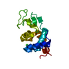 4eteC C: citing same article ( |
|---|---|
| Similar structure data |


-Links
 PDBj
PDBj


- Assembly
| Deposited unit | 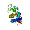
| ||||||||
|---|---|---|---|---|---|---|---|---|---|
| 1 |
| ||||||||
| Unit cell |
| ||||||||
| Components on special symmetry positions |
|
-Components
| #1: Protein | Mass: 14331.160 Da / Num. of mol.: 1 / Source method: isolated from a natural source / Source: (natural) Gallus gallus (chicken) / References: UniProt: P00698, lysozyme |
|---|---|
| #2: Chemical | ChemComp-CL / Chloride  Mass: 35.453 Da / Num. of mol.: 1 / Source method: obtained synthetically / Formula: Cl Mass: 35.453 Da / Num. of mol.: 1 / Source method: obtained synthetically / Formula: Cl |
| #3: Water | ChemComp-HOH / Water Mass: 18.015 Da / Num. of mol.: 73 / Source method: isolated from a natural source / Formula: H2O Mass: 18.015 Da / Num. of mol.: 73 / Source method: isolated from a natural source / Formula: H2O |
-Experimental details
-Experiment
| Experiment | Method: X-RAY DIFFRACTION / Number of used crystals: 1 |
|---|
- Sample preparation
| Crystal | Density Matthews: 2.1 Å3/Da / Density % sol: 41.3 % |
|---|---|
| Crystal grow | Temperature: 293 K / Method: vapor diffusion Details: grown in Linbro plates in hanging or sitting drop geometry. Drops of equal volume of protein (20 mg/ml) and reservoir solution (1 M NaAc pH 3.0, 9-10 % NaCl, 6 % PEG 6000) were mixed. For ...Details: grown in Linbro plates in hanging or sitting drop geometry. Drops of equal volume of protein (20 mg/ml) and reservoir solution (1 M NaAc pH 3.0, 9-10 % NaCl, 6 % PEG 6000) were mixed. For the measurements, the crystals were equilibrated in 1 M NaAc pH 3.4, 10 % NaCl, VAPOR DIFFUSION, temperature 293K |
-Data collection
| Diffraction | Mean temperature: 293 K |
|---|---|
| Diffraction source | Source: SYNCHROTRON / Site: SLS  / Beamline: X10SA / Wavelength: 0.9786 Å / Beamline: X10SA / Wavelength: 0.9786 Å |
| Detector | Type: PSI PILATUS 6M / Detector: PIXEL / Date: Aug 26, 2011 |
| Radiation | Monochromator: SI111 / Protocol: SINGLE WAVELENGTH / Monochromatic (M) / Laue (L): M / Scattering type: x-ray |
| Radiation wavelength | Wavelength: 0.9786 Å / Relative weight: 1 |
| Reflection | Resolution: 1.9→36 Å / Num. all: 10041 / Num. obs: 10041 / % possible obs: 99.9 % / Observed criterion σ(F): 0 / Observed criterion σ(I): 0 |
| Reflection shell | Resolution: 1.9→2 Å / % possible all: 99.9 |
- Processing
| Software |
| |||||||||||||||||||||||||||||||||||
|---|---|---|---|---|---|---|---|---|---|---|---|---|---|---|---|---|---|---|---|---|---|---|---|---|---|---|---|---|---|---|---|---|---|---|---|---|
| Refinement | Method to determine structure: FOURIER SYNTHESIS / Resolution: 1.908→35.464 Å / SU ML: 0.29 / σ(F): 2.01 / Phase error: 16.07 / Stereochemistry target values: ML
| |||||||||||||||||||||||||||||||||||
| Solvent computation | Shrinkage radii: 0.86 Å / VDW probe radii: 1.1 Å / Solvent model: FLAT BULK SOLVENT MODEL / Bsol: 27.125 Å2 / ksol: 0.315 e/Å3 | |||||||||||||||||||||||||||||||||||
| Displacement parameters |
| |||||||||||||||||||||||||||||||||||
| Refinement step | Cycle: LAST / Resolution: 1.908→35.464 Å
| |||||||||||||||||||||||||||||||||||
| Refine LS restraints |
| |||||||||||||||||||||||||||||||||||
| LS refinement shell |
|
