- PDB-4esj: RESTRICTION ENDONUCLEASE DpnI IN COMPLEX WITH TARGET DNA -
+
Open data
ID or keywords:
Loading...
-
Basic information
Entry
Database: PDB / ID: 4esj
Title
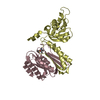
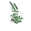
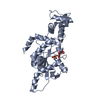
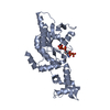
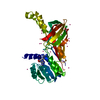
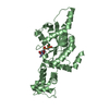
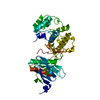
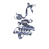
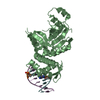
RESTRICTION ENDONUCLEASE DpnI IN COMPLEX WITH TARGET DNA
Components
DNA (5'-D(*CP*TP*GP*GP*(6MA)P*TP*CP*CP*AP*G)-3')
Type-2 restriction enzyme DpnI
Keywords
HYDROLASE/DNA / RESTRICTION ENDONUCLEASE-DNA COMPLEX / TYPE IIM / TYPE IIE / RESTRICTION ENZYME / DPNI / METHYLATION DEPENDENT / N6-METHYLADENINE / PD-(D/E)XK TYPE ENDONUCLEASE / WINGED HELIX DOMAIN / RESTRICTION ENDONUCLEASE / DNA BINDING / HYDROLASE-DNA complex
Function / homology
Function and homology information
type II site-specific deoxyribonuclease / type II site-specific deoxyribonuclease activity / DNA restriction-modification system Similarity search - Function
BIOLOGICAL UNIT CONTAINS A MONOMERIC PROTEIN AND DOUBLE STRANDED DNA FRAGMENT BOUND TO ITS C-TERMINAL WINGED HELIX DOMAIN (ASSEMBLY 1 COMPRISES CHAINS A,C,D; ASSEMBLY 2 COMPRISES CHAINS B,E,F). TO THE BEST OF OUR KNOWLEDGE ADDITIONAL COPY OF DOUBLE STRANDED TARGET DNA BINDS TO THE N-TERMINAL CATALYTIC DOMAIN OF THE ENZYME IN SOLUTION. THE TRIMER (ACCORDING TO PDB CONVENTIONS) IS A COMPLEX OF THE MONOMERIC ENZYME WITH DOUBLE STRANDED DNA FRAGMENT PRESENT IN THE CRYSTAL
-
Components
-
Protein / DNA chain , 2 types, 6 molecules ABCDEF
#1: Protein
Type-2restrictionenzymeDpnI / R.DpnI / Endonuclease DpnI / Type II restriction enzyme DpnI
Mass: 30078.355 Da / Num. of mol.: 2 Source method: isolated from a genetically manipulated source Source: (gene. exp.) Streptococcus pneumoniae (bacteria) / Strain: TIGR4 / Gene: dpnC, spr1665 / Plasmid: PET28 / Production host: Escherichia coli (E. coli) / Strain (production host): ER2925 References: UniProt: P0A460, type II site-specific deoxyribonuclease
#2: DNA chain
DNA (5'-D(*CP*TP*GP*GP*(6MA)P*TP*CP*CP*AP*G)-3')
Mass: 3059.031 Da / Num. of mol.: 4 / Source method: obtained synthetically / Details: synthetic oligonucleotide
Mass: 18.015 Da / Num. of mol.: 197 / Source method: isolated from a natural source / Formula: H2O
-
Experimental details
-
Experiment
Experiment
Method: X-RAY DIFFRACTION / Number of used crystals: 1
-
Sample preparation
Crystal
Density Matthews: 3.44 Å3/Da / Density % sol: 64.29 %
Crystal grow
Temperature: 293 K / Method: vapor diffusion, sitting drop / pH: 6.8 Details: 200 MM POTASSIUM SULFATE, 100 MM BETAINE, 20 % W/V PEG 3350, PH 6.8. FOR CRYOCOOLING THE CRYSTALLIZATION BUFFER WAS MIXED IN 3:1 RATIO WITH 100% GLYCEROL, VAPOR DIFFUSION, SITTING DROP, temperature 293K
Resolution: 2.05→2.1 Å / Redundancy: 3 % / Rmerge(I) obs: 0.3 / Mean I/σ(I) obs: 2.4 / Num. unique all: 4429 / Rsym value: 0.3 / % possible all: 98.4
-
Processing
Software
Name
Version
Classification
ADSC
Quantum
datacollection
SHELXDE
phasing
DM
modelbuilding
ARP/wARP
modelbuilding
REFMAC
5.2.0019
refinement
CNS
refinement
XDS
datareduction
SCALA
datascaling
DM
phasing
Refinement
Method to determine structure: MAD / Resolution: 2.05→19.97 Å / Cor.coef. Fo:Fc: 0.955 / Cor.coef. Fo:Fc free: 0.949 / Cross valid method: THROUGHOUT / ESU R: 0.156 / ESU R Free: 0.137 / Stereochemistry target values: MAXIMUM LIKELIHOOD Details: PROGRAM CNS HAS BEEN USED FOR DNA REFINEMENT. NO SUGAR PUCKER CONSTRAINTS HAVE BEEN APPLIED. HYDROGENS HAVE BEEN ADDED IN THE RIDING POSITIONS. TLS REFINEMENT HAS BEEN USED.
Rfactor
Num. reflection
% reflection
Selection details
Rfree
0.21754
3032
5 %
RANDOM
Rwork
0.19772
-
-
-
all
0.19872
60188
-
-
obs
0.19872
60188
100 %
-
Solvent computation
Ion probe radii: 0.8 Å / Shrinkage radii: 0.8 Å / VDW probe radii: 1.4 Å / Solvent model: MASK
Displacement parameters
Biso mean: 51.616 Å2
Baniso -1
Baniso -2
Baniso -3
1-
0.45 Å2
0 Å2
-0.81 Å2
2-
-
-0.44 Å2
0 Å2
3-
-
-
-0.62 Å2
Refinement step
Cycle: LAST / Resolution: 2.05→19.97 Å
Protein
Nucleic acid
Ligand
Solvent
Total
Num. atoms
4036
730
14
197
4977
Refine LS restraints
Refine-ID
Type
Dev ideal
Dev ideal target
Number
X-RAY DIFFRACTION
r_bond_refined_d
0.011
0.022
5183
X-RAY DIFFRACTION
r_bond_other_d
0
0.02
3449
X-RAY DIFFRACTION
r_angle_refined_deg
1.24
2.134
7150
X-RAY DIFFRACTION
r_angle_other_deg
3.954
3
8425
X-RAY DIFFRACTION
r_dihedral_angle_1_deg
6.112
5
532
X-RAY DIFFRACTION
r_dihedral_angle_2_deg
37.568
24.518
228
X-RAY DIFFRACTION
r_dihedral_angle_3_deg
14.768
15
831
X-RAY DIFFRACTION
r_dihedral_angle_4_deg
19.503
15
28
X-RAY DIFFRACTION
r_chiral_restr
0.077
0.2
753
X-RAY DIFFRACTION
r_gen_planes_refined
0.009
0.02
5311
X-RAY DIFFRACTION
r_gen_planes_other
0.007
0.02
997
X-RAY DIFFRACTION
r_nbd_refined
0.194
0.2
895
X-RAY DIFFRACTION
r_nbd_other
0.246
0.2
3331
X-RAY DIFFRACTION
r_nbtor_refined
0.184
0.2
2423
X-RAY DIFFRACTION
r_nbtor_other
0.109
0.2
2225
X-RAY DIFFRACTION
r_xyhbond_nbd_refined
0.123
0.2
198
X-RAY DIFFRACTION
r_xyhbond_nbd_other
X-RAY DIFFRACTION
r_metal_ion_refined
0.249
0.2
1
X-RAY DIFFRACTION
r_metal_ion_other
X-RAY DIFFRACTION
r_symmetry_vdw_refined
0.183
0.2
18
X-RAY DIFFRACTION
r_symmetry_vdw_other
0.283
0.2
78
X-RAY DIFFRACTION
r_symmetry_hbond_refined
0.181
0.2
25
X-RAY DIFFRACTION
r_symmetry_hbond_other
X-RAY DIFFRACTION
r_symmetry_metal_ion_refined
0.239
0.2
1
X-RAY DIFFRACTION
r_symmetry_metal_ion_other
X-RAY DIFFRACTION
r_mcbond_it
0.657
1.5
2581
X-RAY DIFFRACTION
r_mcbond_other
0
1.5
1032
X-RAY DIFFRACTION
r_mcangle_it
1.201
2
4215
X-RAY DIFFRACTION
r_scbond_it
1.736
3
2602
X-RAY DIFFRACTION
r_scangle_it
2.725
4.5
2935
X-RAY DIFFRACTION
r_rigid_bond_restr
X-RAY DIFFRACTION
r_sphericity_free
X-RAY DIFFRACTION
r_sphericity_bonded
LS refinement shell
Resolution: 2.05→2.103 Å / Total num. of bins used: 20
Rfactor
Num. reflection
% reflection
Rfree
0.305
214
-
Rwork
0.263
4178
-
obs
-
4392
100 %
Refinement TLS params.
Method: refined / Refine-ID: X-RAY DIFFRACTION
ID
L11 (°2)
L12 (°2)
L13 (°2)
L22 (°2)
L23 (°2)
L33 (°2)
S11 (Å °)
S12 (Å °)
S13 (Å °)
S21 (Å °)
S22 (Å °)
S23 (Å °)
S31 (Å °)
S32 (Å °)
S33 (Å °)
T11 (Å2)
T12 (Å2)
T13 (Å2)
T22 (Å2)
T23 (Å2)
T33 (Å2)
Origin x (Å)
Origin y (Å)
Origin z (Å)
1
1.9433
1.4461
0.4369
2.2749
0.2279
0.9341
-0.084
-0.0124
-0.0091
-0.0852
0.0316
-0.0923
-0.1435
0.2281
0.0525
-0.1902
-0.0484
-0.0602
-0.0816
0.0631
0.0579
19.096
20.674
-5.28
2
4.0298
1.7685
0.3665
5.2341
0.3268
2.3816
0.2369
-0.5374
0.0174
0.3173
-0.1256
0.3042
0.3152
-0.3396
-0.1113
-0.1977
-0.1619
-0.0252
0.0019
0.0255
-0.0009
34.629
39.112
9.006
3
1.61
1.3296
1.6435
2.7522
0.9852
3.3425
0.1889
-0.0567
-0.2138
0.4516
0.0526
-0.2363
0.1956
0.159
-0.2415
-0.1062
-0.0012
-0.1
-0.085
-0.0168
0.0492
3.792
10.708
28.681
4
5.7732
1.9756
0.1882
7.2877
2.886
8.0794
0.6462
-0.5814
-0.1607
0.1442
-0.1996
-0.1701
0.3043
0.2283
-0.4466
-0.0147
-0.2056
-0.0573
-0.0616
-0.099
-0.1684
13.031
25.951
50.08
5
2.6037
0.7838
0.1852
3.0645
0.1169
1.7288
0.0307
0.0909
0.0505
0.0308
0.0806
-0.0919
0.1248
-0.1587
-0.1113
-0.1827
-0.0619
-0.0312
-0.044
-0.0615
0.0037
45.3
48.533
2.771
6
2.7575
-0.1684
-1.5812
7.6548
6.3896
9.8561
-0.1073
-0.0045
0.3431
-0.629
0.6961
-0.7602
-0.3177
0.3384
-0.5888
0.0709
-0.3151
0.1144
0.0876
-0.1897
-0.0764
20.27
39.141
52.213
Refinement TLS group
ID
Refine-ID
Refine TLS-ID
Auth asym-ID
Auth seq-ID
1
X-RAY DIFFRACTION
1
A
-1 - 182
2
X-RAY DIFFRACTION
1
A
301
3
X-RAY DIFFRACTION
2
A
183 - 254
4
X-RAY DIFFRACTION
3
B
1 - 182
5
X-RAY DIFFRACTION
3
B
301
6
X-RAY DIFFRACTION
4
B
183 - 254
7
X-RAY DIFFRACTION
5
C
1 - 10
8
X-RAY DIFFRACTION
5
D
1 - 10
9
X-RAY DIFFRACTION
6
E
3 - 10
10
X-RAY DIFFRACTION
6
F
1 - 8
+
About Yorodumi
-
News
-
Feb 9, 2022. New format data for meta-information of EMDB entries
New format data for meta-information of EMDB entries
Version 3 of the EMDB header file is now the official format.
The previous official version 1.9 will be removed from the archive.
In the structure databanks used in Yorodumi, some data are registered as the other names, "COVID-19 virus" and "2019-nCoV". Here are the details of the virus and the list of structure data.
Jan 31, 2019. EMDB accession codes are about to change! (news from PDBe EMDB page)
EMDB accession codes are about to change! (news from PDBe EMDB page)
The allocation of 4 digits for EMDB accession codes will soon come to an end. Whilst these codes will remain in use, new EMDB accession codes will include an additional digit and will expand incrementally as the available range of codes is exhausted. The current 4-digit format prefixed with “EMD-” (i.e. EMD-XXXX) will advance to a 5-digit format (i.e. EMD-XXXXX), and so on. It is currently estimated that the 4-digit codes will be depleted around Spring 2019, at which point the 5-digit format will come into force.
The EM Navigator/Yorodumi systems omit the EMD- prefix.
Related info.:Q: What is EMD? / ID/Accession-code notation in Yorodumi/EM Navigator
Yorodumi is a browser for structure data from EMDB, PDB, SASBDB, etc.
This page is also the successor to EM Navigator detail page, and also detail information page/front-end page for Omokage search.
The word "yorodu" (or yorozu) is an old Japanese word meaning "ten thousand". "mi" (miru) is to see.
Related info.:EMDB / PDB / SASBDB / Comparison of 3 databanks / Yorodumi Search / Aug 31, 2016. New EM Navigator & Yorodumi / Yorodumi Papers / Jmol/JSmol / Function and homology information / Changes in new EM Navigator and Yorodumi
 Movie
Movie Controller


 Open data
 Basic information
 Components
 Keywords
 RESTRICTION ENZYME / DPNI / METHYLATION DEPENDENT /
  Function and homology information

 Authors
 Citation
 Structure visualization
 Downloads & links
 Other downloads
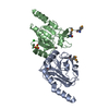

 PDBj
PDBj

 Assembly
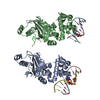


 Mass: 65.409 Da / Num. of mol.: 3 / Source method: obtained synthetically / Formula: Zn
 Mass: 42.020 Da / Num. of mol.: 1 / Source method: obtained synthetically / Formula: N3
 Num. of mol.: 2 / Source method: obtained synthetically
 Mass: 92.094 Da / Num. of mol.: 1 / Source method: obtained synthetically / Formula: C3H8O3
 Sample preparation
 / Beamline: I02 / Wavelength: 0.97949,1.2833,1.2835
 Processing