- PDB-4drb: The crystal structure of FANCM bound MHF complex -
+
Open data
ID or keywords:
Loading...
-
Basic information
Entry
Database: PDB / ID: 4drb
Title
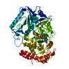
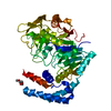
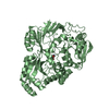
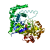
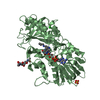
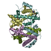
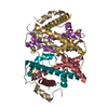
The crystal structure of FANCM bound MHF complex
Components
Centromere protein S
Centromere protein X
Fanconi anemia group M protein
Keywords
DNA BINDING PROTEIN/PROTEIN BINDING / DNA repair / DNA binding complex / Histone fold / DNA damage repair / DNA binding / DNA BINDING-PROTEIN BINDING complex / DNA BINDING PROTEIN-PROTEIN BINDING complex
Function / homology
Function and homology information
double-strand break repair via synthesis-dependent strand annealing / FANCM-MHF complex / Fanconi anaemia nuclear complex / inner kinetochore / four-way junction helicase activity / resolution of meiotic recombination intermediates / nuclease activity / kinetochore assembly / 3'-5' DNA helicase activity / positive regulation of protein monoubiquitination ...double-strand break repair via synthesis-dependent strand annealing / FANCM-MHF complex / Fanconi anaemia nuclear complex / inner kinetochore / four-way junction helicase activity / resolution of meiotic recombination intermediates / nuclease activity / kinetochore assembly / 3'-5' DNA helicase activity / positive regulation of protein monoubiquitination / replication fork processing / Amplification of signal from unattached kinetochores via a MAD2 inhibitory signal / interstrand cross-link repair / Mitotic Prometaphase / four-way junction DNA binding / EML4 and NUDC in mitotic spindle formation / Deposition of new CENPA-containing nucleosomes at the centromere / Resolution of Sister Chromatid Cohesion / positive regulation of protein ubiquitination / chromosome segregation / RHO GTPases Activate Formins / Fanconi Anemia Pathway / PKR-mediated signaling / Separation of Sister Chromatids / RNA helicase activity / RNA helicase / protein heterodimerization activity / cell division / DNA repair / DNA damage response / chromatin binding / chromatin / ATP hydrolysis activity / DNA binding / nucleoplasm / ATP binding / nucleus / cytosol Similarity search - Function
FANCM, DEAH-box helicase domain / : / Fanconi anemia group M protein, MHF binding domain / FANCM/Mph1-like / FANCM to MHF binding domain / GTP Cyclohydrolase I; Chain A, domain 1 - #30 / Single alpha-helices involved in coiled-coils or other helix-helix interfaces - #4980 / GTP Cyclohydrolase I; Chain A, domain 1 / Centromere protein X / CENP-S/Mhf1 ...FANCM, DEAH-box helicase domain / : / Fanconi anemia group M protein, MHF binding domain / FANCM/Mph1-like / FANCM to MHF binding domain / GTP Cyclohydrolase I; Chain A, domain 1 - #30 / Single alpha-helices involved in coiled-coils or other helix-helix interfaces - #4980 / GTP Cyclohydrolase I; Chain A, domain 1 / Centromere protein X / CENP-S/Mhf1 / CENP-S associating Centromere protein X / CENP-S protein / ERCC4 domain / ERCC4 domain / ERCC4 domain / RuvA domain 2-like / Restriction endonuclease type II-like / Histone, subunit A / Histone, subunit A / DEAD/DEAH box helicase / DEAD/DEAH box helicase domain / Single alpha-helices involved in coiled-coils or other helix-helix interfaces / Helix non-globular / Helicase conserved C-terminal domain / Special / Histone-fold / helicase superfamily c-terminal domain / Superfamilies 1 and 2 helicase C-terminal domain profile. / Superfamilies 1 and 2 helicase ATP-binding type-1 domain profile. / DEAD-like helicases superfamily / Helicase, C-terminal / Helicase superfamily 1/2, ATP-binding domain / Up-down Bundle / P-loop containing nucleoside triphosphate hydrolase / Orthogonal Bundle / Mainly Alpha Similarity search - Domain/homology
A: Centromere protein S B: Centromere protein S C: Fanconi anemia group M protein D: Centromere protein S E: Centromere protein S F: Fanconi anemia group M protein G: Centromere protein S H: Centromere protein S I: Fanconi anemia group M protein J: Centromere protein X K: Centromere protein X L: Centromere protein X M: Centromere protein X N: Centromere protein X O: Centromere protein X
In the structure databanks used in Yorodumi, some data are registered as the other names, "COVID-19 virus" and "2019-nCoV". Here are the details of the virus and the list of structure data.
Jan 31, 2019. EMDB accession codes are about to change! (news from PDBe EMDB page)
EMDB accession codes are about to change! (news from PDBe EMDB page)
The allocation of 4 digits for EMDB accession codes will soon come to an end. Whilst these codes will remain in use, new EMDB accession codes will include an additional digit and will expand incrementally as the available range of codes is exhausted. The current 4-digit format prefixed with “EMD-” (i.e. EMD-XXXX) will advance to a 5-digit format (i.e. EMD-XXXXX), and so on. It is currently estimated that the 4-digit codes will be depleted around Spring 2019, at which point the 5-digit format will come into force.
The EM Navigator/Yorodumi systems omit the EMD- prefix.
Related info.:Q: What is EMD? / ID/Accession-code notation in Yorodumi/EM Navigator
Yorodumi is a browser for structure data from EMDB, PDB, SASBDB, etc.
This page is also the successor to EM Navigator detail page, and also detail information page/front-end page for Omokage search.
The word "yorodu" (or yorozu) is an old Japanese word meaning "ten thousand". "mi" (miru) is to see.
Related info.:EMDB / PDB / SASBDB / Comparison of 3 databanks / Yorodumi Search / Aug 31, 2016. New EM Navigator & Yorodumi / Yorodumi Papers / Jmol/JSmol / Function and homology information / Changes in new EM Navigator and Yorodumi
 Movie
Movie Controller


 Open data
 Basic information
 Components
 Keywords
 DNA repair / DNA binding complex /
  Function and homology information
 Authors
 Citation
 Structure visualization
 Downloads & links
 Other downloads


 PDBj
PDBj


 Assembly
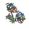


 Mass: 18.015 Da / Num. of mol.: 120 / Source method: isolated from a natural source / Formula: H2O
 Sample preparation
 / Beamline: BL17U / Wavelength: 0.9792 Å
 Processing