 Movie
Movie Controller

[English] 日本語
 Yorodumi
- PDB-4dbz: Crystal Structure of V151L Actinorhodin Polyketide Ketoreductase ... -
+ Open data

- Basic information
| Entry | Database: PDB / ID: 4dbz | ||||||
|---|---|---|---|---|---|---|---|
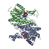
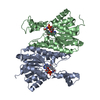
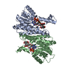
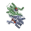
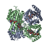
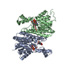
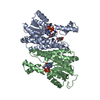
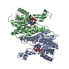
| Title | Crystal Structure of V151L Actinorhodin Polyketide Ketoreductase with NADPH | ||||||
 Components Components | Ketoacyl reductase | ||||||
 Keywords Keywords |  OXIDOREDUCTASE / Rossmann fold / ketoreductase OXIDOREDUCTASE / Rossmann fold / ketoreductase | ||||||
| Function / homology |  Function and homology informationOxidoreductases; Acting on the CH-CH group of donors; With NAD+ or NADP+ as acceptor / steroid metabolic process / antibiotic biosynthetic process / oxidoreductase activity Function and homology informationOxidoreductases; Acting on the CH-CH group of donors; With NAD+ or NADP+ as acceptor / steroid metabolic process / antibiotic biosynthetic process / oxidoreductase activitySimilarity search - Function | ||||||
| Biological species |  Streptomyces coelicolor (bacteria) Streptomyces coelicolor (bacteria) | ||||||
| Method | X-RAY DIFFRACTION / SYNCHROTRON / MOLECULAR REPLACEMENT / Resolution: 2.643 Å | ||||||
 Authors Authors | Javidpour, P. / Tsai, S.-C. | ||||||
 Citation Citation | Journal: Chem.Biol. / Year: 2013 Title: The Determinants of Activity and Specificity in Actinorhodin Type II Polyketide Ketoreductase. Authors: Javidpour, P. / Bruegger, J. / Srithahan, S. / Korman, T.P. / Crump, M.P. / Crosby, J. / Burkart, M.D. / Tsai, S.C. | ||||||
| History |
|
- Structure visualization
| Structure viewer | Molecule: MolmilJmol/JSmol |
|---|
- Downloads & links
-Download
| PDBx/mmCIF format | 4dbz.cif.gz | 108.2 KB | Display | PDBx/mmCIF format |
|---|---|---|---|---|
| PDB format | pdb4dbz.ent.gz | 84.1 KB | Display | PDB format |
| PDBx/mmJSON format | 4dbz.json.gz | Tree view | PDBx/mmJSON format | |
| Others |  Other downloads Other downloads |
-Validation report
| Arichive directory | https://data.pdbj.org/pub/pdb/validation_reports/db/4dbzftp://data.pdbj.org/pub/pdb/validation_reports/db/4dbz | HTTPS FTP |
|---|
-Related structure data


-Links
 PDBj
PDBj


- Assembly
| Deposited unit | 
| ||||||||
|---|---|---|---|---|---|---|---|---|---|
| 1 | 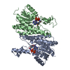
| ||||||||
| Unit cell |
|
-Components
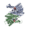
| #1: Protein | Mass: 29478.389 Da / Num. of mol.: 2 / Mutation: V151L Source method: isolated from a genetically manipulated source Source: (gene. exp.) Streptomyces coelicolor (bacteria) / Gene: actIII, SCBAC28G1.12c, SCO5086 / Plasmid: pET28 / Production host: Escherichia coli (E. coli) / Strain (production host): BL21(DE3)References: UniProt: P16544, Oxidoreductases; Acting on the CH-CH group of donors; With NAD+ or NADP+ as acceptor#2: Chemical | Nicotinamide adenine dinucleotide phosphate  Mass: 745.421 Da / Num. of mol.: 2 / Source method: obtained synthetically / Formula: C21H30N7O17P3 Mass: 745.421 Da / Num. of mol.: 2 / Source method: obtained synthetically / Formula: C21H30N7O17P3#3: Water | ChemComp-HOH / | Water Mass: 18.015 Da / Num. of mol.: 44 / Source method: isolated from a natural source / Formula: H2O Mass: 18.015 Da / Num. of mol.: 44 / Source method: isolated from a natural source / Formula: H2O |
|---|
-Experimental details
-Experiment
| Experiment | Method: X-RAY DIFFRACTION / Number of used crystals: 1 |
|---|
- Sample preparation
| Crystal | Density Matthews: 3.16 Å3/Da / Density % sol: 61.08 % |
|---|---|
| Crystal grow | Temperature: 298 K / Method: vapor diffusion, sitting drop / pH: 9 Details: 0.1 M Tris-Cl, 1.8 M Na malonate, pH 9.0, VAPOR DIFFUSION, SITTING DROP, temperature 298K |
-Data collection
| Diffraction | Mean temperature: 100 K |
|---|---|
| Diffraction source | Source: SYNCHROTRON / Site: ALS  / Beamline: 8.2.2 / Wavelength: 0.9998 Å / Beamline: 8.2.2 / Wavelength: 0.9998 Å |
| Detector | Type: ADSC QUANTUM 315 / Detector: CCD / Date: Feb 25, 2009 |
| Radiation | Protocol: SINGLE WAVELENGTH / Monochromatic (M) / Laue (L): M / Scattering type: x-ray |
| Radiation wavelength | Wavelength: 0.9998 Å / Relative weight: 1 |
| Reflection | Resolution: 2.64→50 Å / Num. obs: 21581 |
- Processing
| Software |
| |||||||||||||||||||||||||||||||||||||||||||||||||||||||||||||||
|---|---|---|---|---|---|---|---|---|---|---|---|---|---|---|---|---|---|---|---|---|---|---|---|---|---|---|---|---|---|---|---|---|---|---|---|---|---|---|---|---|---|---|---|---|---|---|---|---|---|---|---|---|---|---|---|---|---|---|---|---|---|---|---|---|
| Refinement | Method to determine structure: MOLECULAR REPLACEMENT / Resolution: 2.643→47.4185 Å / SU ML: 0.3 / σ(F): 0 / Phase error: 21.04 / Stereochemistry target values: ML
| |||||||||||||||||||||||||||||||||||||||||||||||||||||||||||||||
| Solvent computation | Shrinkage radii: 0.83 Å / VDW probe radii: 1.1 Å / Solvent model: FLAT BULK SOLVENT MODEL / Bsol: 43.308 Å2 / ksol: 0.401 e/Å3 | |||||||||||||||||||||||||||||||||||||||||||||||||||||||||||||||
| Displacement parameters |
| |||||||||||||||||||||||||||||||||||||||||||||||||||||||||||||||
| Refinement step | Cycle: LAST / Resolution: 2.643→47.4185 Å
| |||||||||||||||||||||||||||||||||||||||||||||||||||||||||||||||
| Refine LS restraints |
| |||||||||||||||||||||||||||||||||||||||||||||||||||||||||||||||
| LS refinement shell |
|
