 Movie
Movie Controller

[English] 日本語
 Yorodumi
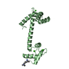
- PDB-4cln: STRUCTURE OF A RECOMBINANT CALMODULIN FROM DROSOPHILA MELANOGASTE... -
+ Open data

- Basic information
| Entry | Database: PDB / ID: 4cln | ||||||
|---|---|---|---|---|---|---|---|
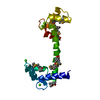
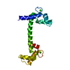
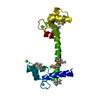
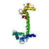
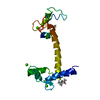
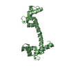
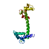
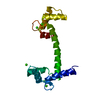
| Title | STRUCTURE OF A RECOMBINANT CALMODULIN FROM DROSOPHILA MELANOGASTER REFINED AT 2.2-ANGSTROMS RESOLUTION | ||||||
 Components Components | CALMODULIN | ||||||
 Keywords Keywords | CALCIUM BINDING PROTEIN | ||||||
| Function / homology |  Function and homology information Function and homology informationmetarhodopsin inactivation / myosin VI complex / myosin VI head/neck binding / myosin VII complex / photoreceptor cell axon guidance / negative regulation of opsin-mediated signaling pathway / rhabdomere / rhabdomere development / myosin V complex / : ...metarhodopsin inactivation / myosin VI complex / myosin VI head/neck binding / myosin VII complex / photoreceptor cell axon guidance / negative regulation of opsin-mediated signaling pathway / rhabdomere / rhabdomere development / myosin V complex / : / kinetochore organization / : / actin filament-based movement / rhodopsin mediated signaling pathway / Neutrophil degranulation / myosin V binding / channel regulator activity / cellular response to ethanol / muscle cell cellular homeostasis / myosin heavy chain binding / mitotic spindle pole / centriole replication / enzyme regulator activity / centriole / sensory perception of sound / spindle / mitotic spindle / sensory perception of smell / cell cortex / midbody / protein phosphorylation / centrosome / calcium ion binding / nucleoplasm / cytosol / cytoplasmSimilarity search - Function | ||||||
| Biological species |  Drosophila melanogaster (fruit fly) Drosophila melanogaster (fruit fly) | ||||||
| Method | X-RAY DIFFRACTION / Resolution: 2.2 Å | ||||||
 Authors Authors | Taylor, D.A. / Sack, J.S. / Maune, J.F. / Beckingham, K. / Quiocho, F.A. | ||||||
 Citation Citation | Journal: J.Biol.Chem. / Year: 1991 Title: Structure of a recombinant calmodulin from Drosophila melanogaster refined at 2.2-A resolution. Authors: Taylor, D.A. / Sack, J.S. / Maune, J.F. / Beckingham, K. / Quiocho, F.A. #1: Journal: J.Mol.Biol. / Year: 1987Title: Structure and Sequence of the Drosophila Melanogaster Calmodulin Gene Authors: Smith, V.L. / Doyle, K.E. / Maune, J.F. / Munjaal, R.P. / Beckingham, K. | ||||||
| History |
|
- Structure visualization
| Structure viewer | Molecule: MolmilJmol/JSmol |
|---|
- Downloads & links
-Download
| PDBx/mmCIF format | 4cln.cif.gz | 44.2 KB | Display | PDBx/mmCIF format |
|---|---|---|---|---|
| PDB format | pdb4cln.ent.gz | 30.9 KB | Display | PDB format |
| PDBx/mmJSON format | 4cln.json.gz | Tree view | PDBx/mmJSON format | |
| Others |  Other downloads Other downloads |
-Validation report
| Arichive directory | https://data.pdbj.org/pub/pdb/validation_reports/cl/4clnftp://data.pdbj.org/pub/pdb/validation_reports/cl/4cln | HTTPS FTP |
|---|
-Related structure data
| Similar structure data |
|---|

-Links
 PDBj
PDBj

- Assembly
| Deposited unit | 
| ||||||||
|---|---|---|---|---|---|---|---|---|---|
| 1 |
| ||||||||
| Unit cell |
|
-Components
| #1: Protein | Mass: 16694.324 Da / Num. of mol.: 1 Source method: isolated from a genetically manipulated source Source: (gene. exp.) Drosophila melanogaster (fruit fly) / References: UniProt: P62152 | ||
|---|---|---|---|
| #2: Chemical | ChemComp-CA /   Mass: 40.078 Da / Num. of mol.: 4 / Source method: obtained synthetically / Formula: Ca Mass: 40.078 Da / Num. of mol.: 4 / Source method: obtained synthetically / Formula: Ca#3: Water | ChemComp-HOH / | Water Mass: 18.015 Da / Num. of mol.: 78 / Source method: isolated from a natural source / Formula: H2O Mass: 18.015 Da / Num. of mol.: 78 / Source method: isolated from a natural source / Formula: H2O |
-Experimental details
-Experiment
| Experiment | Method: X-RAY DIFFRACTION |
|---|
- Sample preparation
| Crystal | Density Matthews: 2.34 Å3/Da / Density % sol: 47.53 % | ||||||||||||||||||||||||||||||
|---|---|---|---|---|---|---|---|---|---|---|---|---|---|---|---|---|---|---|---|---|---|---|---|---|---|---|---|---|---|---|---|
| Crystal grow | *PLUS Temperature: 18 ℃ / Method: vapor diffusion, hanging drop / pH: 4 / Details: initial seeding crystal | ||||||||||||||||||||||||||||||
| Components of the solutions | *PLUS
|
-Data collection
| Reflection | *PLUS Highest resolution: 2.2 Å / Lowest resolution: 20 Å / Num. obs: 6831 / % possible obs: 81.7 % / Observed criterion σ(F): 2 / Num. measured all: 10617 / Rmerge F obs: 0.068 |
|---|
- Processing
| Software | Name: PROLSQ / Classification: refinement | ||||||||||||||||||||||||||||||||||||||||||||||||||||||||||||||||||||||||||||||||||||
|---|---|---|---|---|---|---|---|---|---|---|---|---|---|---|---|---|---|---|---|---|---|---|---|---|---|---|---|---|---|---|---|---|---|---|---|---|---|---|---|---|---|---|---|---|---|---|---|---|---|---|---|---|---|---|---|---|---|---|---|---|---|---|---|---|---|---|---|---|---|---|---|---|---|---|---|---|---|---|---|---|---|---|---|---|---|
| Refinement | Resolution: 2.2→10 Å / σ(F): 2 /
| ||||||||||||||||||||||||||||||||||||||||||||||||||||||||||||||||||||||||||||||||||||
| Refinement step | Cycle: LAST / Resolution: 2.2→10 Å
| ||||||||||||||||||||||||||||||||||||||||||||||||||||||||||||||||||||||||||||||||||||
| Refine LS restraints |
| ||||||||||||||||||||||||||||||||||||||||||||||||||||||||||||||||||||||||||||||||||||
| Software | *PLUS Name: 'X-PLOR, PROLSQ' / Classification: refinement | ||||||||||||||||||||||||||||||||||||||||||||||||||||||||||||||||||||||||||||||||||||
| Refinement | *PLUS Highest resolution: 2.2 Å / Lowest resolution: 10 Å / Num. reflection obs: 5239 / σ(F): 2 / Rfactor obs: 0.197 | ||||||||||||||||||||||||||||||||||||||||||||||||||||||||||||||||||||||||||||||||||||
| Solvent computation | *PLUS | ||||||||||||||||||||||||||||||||||||||||||||||||||||||||||||||||||||||||||||||||||||
| Displacement parameters | *PLUS |
