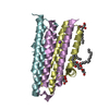
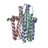
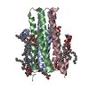
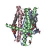
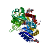
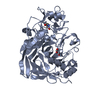
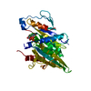
Journal: To be Published Title: Crystal Structure of the Integral Membrane Diacylglycerol Kinase with Zn-Amppcp Bound and its Catalytic Mechanism Authors: Li, D. / Caffrey, M.
History
Deposition
Dec 23, 2013
Deposition site: PDBE / Processing site: PDBE
Revision 1.0
Jan 28, 2015
Provider: repository / Type: Initial release
Revision 1.1
Nov 16, 2016
Group: Database references
Revision 1.2
Mar 6, 2019
Group: Data collection / Experimental preparation / Other Category: exptl_crystal_grow / pdbx_database_proc / pdbx_database_status Item: _exptl_crystal_grow.method / _pdbx_database_status.recvd_author_approval
Mass: 18.015 Da / Num. of mol.: 1 / Source method: isolated from a natural source / Formula: H2O
Sequence details
THE CONSTRUCT CONTAINS AN N-TERMIANL HIS TAG 'GHHHHHHEL'. COMPARED TO THE WILDTYPE FORM, THE ...THE CONSTRUCT CONTAINS AN N-TERMIANL HIS TAG 'GHHHHHHEL'. COMPARED TO THE WILDTYPE FORM, THE PROTEIN HAS FOUR MUTATIONS. THEY ARE I53C, I70L, M96L AND V107D.
-
Experimental details
-
Experiment
Experiment
Method: X-RAY DIFFRACTION / Number of used crystals: 1
-
Sample preparation
Crystal
Density Matthews: 3.57 Å3/Da / Density % sol: 65.53 % / Description: NONE
Crystal grow
Temperature: 277 K / Method: lipidic cubic phase / pH: 5.6 Details: 7-9%(V/V) 2-METHYL-2-4-PENTANEDIOL, 0.1 M SODIUM CHLORIDE, 0.1 M LITHIUM NITRATE, 0.1 M SODIUM CITRATE PH 5.6. CRYSTALLIZED USING THE IN MESO (LIPIDIC CUBIC PHASE) METHOD AT 4 DEGREE CELSIUS ...Details: 7-9%(V/V) 2-METHYL-2-4-PENTANEDIOL, 0.1 M SODIUM CHLORIDE, 0.1 M LITHIUM NITRATE, 0.1 M SODIUM CITRATE PH 5.6. CRYSTALLIZED USING THE IN MESO (LIPIDIC CUBIC PHASE) METHOD AT 4 DEGREE CELSIUS WITH THE MONOOLEIN AS THE HOSTING LIPID. CRYSTALS WERE SOAKED AT 4 DEGREE CELSIUS WITH 10 MM AMPPCP AND 60 MM MAGNESIUM IN THE CRYSTALLIZATION CONDITION FOR 2 H BEFORE HARVESTING.
In the structure databanks used in Yorodumi, some data are registered as the other names, "COVID-19 virus" and "2019-nCoV". Here are the details of the virus and the list of structure data.
Jan 31, 2019. EMDB accession codes are about to change! (news from PDBe EMDB page)
EMDB accession codes are about to change! (news from PDBe EMDB page)
The allocation of 4 digits for EMDB accession codes will soon come to an end. Whilst these codes will remain in use, new EMDB accession codes will include an additional digit and will expand incrementally as the available range of codes is exhausted. The current 4-digit format prefixed with “EMD-” (i.e. EMD-XXXX) will advance to a 5-digit format (i.e. EMD-XXXXX), and so on. It is currently estimated that the 4-digit codes will be depleted around Spring 2019, at which point the 5-digit format will come into force.
The EM Navigator/Yorodumi systems omit the EMD- prefix.
Related info.:Q: What is EMD? / ID/Accession-code notation in Yorodumi/EM Navigator
Yorodumi is a browser for structure data from EMDB, PDB, SASBDB, etc.
This page is also the successor to EM Navigator detail page, and also detail information page/front-end page for Omokage search.
The word "yorodu" (or yorozu) is an old Japanese word meaning "ten thousand". "mi" (miru) is to see.
Related info.:EMDB / PDB / SASBDB / Comparison of 3 databanks / Yorodumi Search / Aug 31, 2016. New EM Navigator & Yorodumi / Yorodumi Papers / Jmol/JSmol / Function and homology information / Changes in new EM Navigator and Yorodumi
 Movie
Movie Controller

 Yorodumi
 Open data
 Basic information
 Components
 Keywords
 Function and homology information
 Authors
 Citation
 Structure visualization
 Downloads & links
 Other downloads
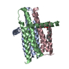


 PDBj
PDBj


 Assembly

 Mass: 356.540 Da / Num. of mol.: 2 / Source method: obtained synthetically / Formula: C21H40O4
 Mass: 65.409 Da / Num. of mol.: 2 / Source method: obtained synthetically / Formula: Zn
 Mass: 505.208 Da / Num. of mol.: 1 / Source method: obtained synthetically / Formula: C11H18N5O12P3 / Comment: AMP-PCP, energy-carrying molecule analogue*YM
 Mass: 18.015 Da / Num. of mol.: 1 / Source method: isolated from a natural source / Formula: H2O
 Sample preparation
 / Beamline: 23-ID-B / Wavelength: 1.0332
  Processing