 Movie
Movie Controller

[English] 日本語
 Yorodumi
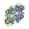
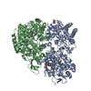
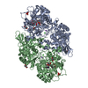
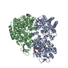
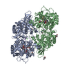
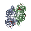
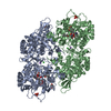
- PDB-4c50: Crystal Structure of the Catalase-Peroxidase (KatG) D137S mutant ... -
+ Open data

- Basic information
| Entry | Database: PDB / ID: 4c50 | ||||||
|---|---|---|---|---|---|---|---|
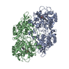
| Title | Crystal Structure of the Catalase-Peroxidase (KatG) D137S mutant from Mycobacterium Tuberculosis | ||||||
 Components Components | CATALASE-PEROXIDASE | ||||||
 Keywords Keywords | OXIDOREDUCTASE | ||||||
| Function / homology |  Function and homology information Function and homology informationoxidoreductase activity, acting on a heme group of donors, nitrogenous group as acceptor / Tolerance of reactive oxygen produced by macrophages / catalase-peroxidase / NADH binding / cell wall / catalase activity / NADPH binding / positive regulation of DNA repair / peroxidase activity / peptidoglycan-based cell wall ...oxidoreductase activity, acting on a heme group of donors, nitrogenous group as acceptor / Tolerance of reactive oxygen produced by macrophages / catalase-peroxidase / NADH binding / cell wall / catalase activity / NADPH binding / positive regulation of DNA repair / peroxidase activity / peptidoglycan-based cell wall / hydrogen peroxide catabolic process / cellular response to hydrogen peroxide / response to oxidative stress / response to antibiotic / heme binding / extracellular region / metal ion binding / plasma membrane / cytosolSimilarity search - Function | ||||||
| Biological species |   MYCOBACTERIUM TUBERCULOSIS (bacteria) MYCOBACTERIUM TUBERCULOSIS (bacteria) | ||||||
| Method | X-RAY DIFFRACTION / SYNCHROTRON / MOLECULAR REPLACEMENT / Resolution: 2.5 Å | ||||||
 Authors Authors | Hersleth, H.-P. / Zhao, X. / Magliozzo, R.S. / Andersson, K.K. | ||||||
 Citation Citation | Journal: Chem.Commun.(Camb.) / Year: 2013 Title: Access Channel Residues Ser315 and Asp137 in Mycobacterium Tuberculosis Catalase-Peroxidase (Katg) Control Peroxidatic Activation of the Pro-Drug Isoniazid. Authors: Zhao, X. / Hersleth, H.P. / Zhu, J. / Andersson, K.K. / Magliozzo, R.S. | ||||||
| History |
|
- Structure visualization
| Structure viewer | Molecule: MolmilJmol/JSmol |
|---|
- Downloads & links
-Download
| PDBx/mmCIF format | 4c50.cif.gz | 283.1 KB | Display | PDBx/mmCIF format |
|---|---|---|---|---|
| PDB format | pdb4c50.ent.gz | 228.6 KB | Display | PDB format |
| PDBx/mmJSON format | 4c50.json.gz | Tree view | PDBx/mmJSON format | |
| Others |  Other downloads Other downloads |
-Validation report
| Arichive directory | https://data.pdbj.org/pub/pdb/validation_reports/c5/4c50ftp://data.pdbj.org/pub/pdb/validation_reports/c5/4c50 | HTTPS FTP |
|---|
-Related structure data
| Related structure data |  4c51C 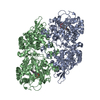 2ccaS C: citing same article ( S: Starting model for refinement |
|---|---|
| Similar structure data |


-Links
 PDBj
PDBj


- Assembly
| Deposited unit | 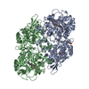
| ||||||||||||||||||||||||||||||
|---|---|---|---|---|---|---|---|---|---|---|---|---|---|---|---|---|---|---|---|---|---|---|---|---|---|---|---|---|---|---|---|
| 1 |
| ||||||||||||||||||||||||||||||
| Unit cell |
| ||||||||||||||||||||||||||||||
| Noncrystallographic symmetry (NCS) | NCS domain:
NCS domain segments: Component-ID: 1 / Ens-ID: 1 / Beg auth comp-ID: HIS / Beg label comp-ID: HIS / End auth comp-ID: ARG / End label comp-ID: ARG / Refine code: 1 / Auth seq-ID: 25 - 740 / Label seq-ID: 25 - 740
NCS oper:
|
-Components
| #1: Protein | / CP / PEROXIDASE/CATALASE Mass: 80659.602 Da / Num. of mol.: 2 / Mutation: YES Source method: isolated from a genetically manipulated source Details: MYW-ADDUCT BETWEEN M255 Y229 W107 / Source: (gene. exp.) MYCOBACTERIUM TUBERCULOSIS (bacteria) / Strain: H37RV / Production host: ESCHERICHIA COLI (E. coli)References: UniProt: Q08129, UniProt: P9WIE5*PLUS, catalase-peroxidase#2: Sugar | ChemComp-GLC / | Glucose  Type: D-saccharide, alpha linking / Mass: 180.156 Da / Num. of mol.: 1 Type: D-saccharide, alpha linking / Mass: 180.156 Da / Num. of mol.: 1Source method: isolated from a genetically manipulated source Formula: C6H12O6 #3: Chemical | Heme B  Mass: 616.487 Da / Num. of mol.: 2 / Source method: obtained synthetically / Formula: C34H32FeN4O4 Mass: 616.487 Da / Num. of mol.: 2 / Source method: obtained synthetically / Formula: C34H32FeN4O4#4: Chemical | ChemComp-ACT / | Acetate  Mass: 59.044 Da / Num. of mol.: 1 / Source method: obtained synthetically / Formula: C2H3O2 Mass: 59.044 Da / Num. of mol.: 1 / Source method: obtained synthetically / Formula: C2H3O2#5: Water | ChemComp-HOH / | Water Mass: 18.015 Da / Num. of mol.: 82 / Source method: isolated from a natural source / Formula: H2O Mass: 18.015 Da / Num. of mol.: 82 / Source method: isolated from a natural source / Formula: H2O |
|---|
-Experimental details
-Experiment
| Experiment | Method: X-RAY DIFFRACTION / Number of used crystals: 1 |
|---|
- Sample preparation
| Crystal | Density Matthews: 2.83 Å3/Da / Density % sol: 56.3 % / Description: NONE |
|---|---|
| Crystal grow | pH: 4.6 Details: 100 MM SODIUM ACETATE, PH 4.6, 6% PEG 4000, AND 0.17 MM N-DODECYL-BETA-D-MALTOSIDE |
-Data collection
| Diffraction | Mean temperature: 100 K |
|---|---|
| Diffraction source | Source: SYNCHROTRON / Site: SLS  / Beamline: X10SA / Wavelength: 1 / Beamline: X10SA / Wavelength: 1 |
| Detector | Type: DECTRIS PILATUS 6M / Detector: PIXEL / Date: Nov 30, 2011 |
| Radiation | Protocol: SINGLE WAVELENGTH / Monochromatic (M) / Laue (L): M / Scattering type: x-ray |
| Radiation wavelength | Wavelength: 1 Å / Relative weight: 1 |
| Reflection | Resolution: 2.5→75.3 Å / Num. obs: 62942 / % possible obs: 99.9 % / Observed criterion σ(I): 6 / Redundancy: 6.2 % / Rmerge(I) obs: 0.13 / Net I/σ(I): 8.7 |
| Reflection shell | Resolution: 2.5→2.56 Å / Redundancy: 6.5 % / Rmerge(I) obs: 0.39 / Mean I/σ(I) obs: 3.9 / % possible all: 100 |
- Processing
| Software |
| ||||||||||||||||||||||||||||||||||||||||||||||||||||||||||||||||||||||||||||||||||||||||||||||||||||||||||||||||||||||||||||||||||||||||||||||||||||||||||||||||||||||||||||||||||||||
|---|---|---|---|---|---|---|---|---|---|---|---|---|---|---|---|---|---|---|---|---|---|---|---|---|---|---|---|---|---|---|---|---|---|---|---|---|---|---|---|---|---|---|---|---|---|---|---|---|---|---|---|---|---|---|---|---|---|---|---|---|---|---|---|---|---|---|---|---|---|---|---|---|---|---|---|---|---|---|---|---|---|---|---|---|---|---|---|---|---|---|---|---|---|---|---|---|---|---|---|---|---|---|---|---|---|---|---|---|---|---|---|---|---|---|---|---|---|---|---|---|---|---|---|---|---|---|---|---|---|---|---|---|---|---|---|---|---|---|---|---|---|---|---|---|---|---|---|---|---|---|---|---|---|---|---|---|---|---|---|---|---|---|---|---|---|---|---|---|---|---|---|---|---|---|---|---|---|---|---|---|---|---|---|
| Refinement | Method to determine structure: MOLECULAR REPLACEMENT Starting model: PDB ENTRY 2CCA Resolution: 2.5→75.41 Å / Cor.coef. Fo:Fc: 0.876 / Cor.coef. Fo:Fc free: 0.824 / SU B: 15.465 / SU ML: 0.313 / Cross valid method: THROUGHOUT / ESU R: 0.587 / ESU R Free: 0.344 / Stereochemistry target values: MAXIMUM LIKELIHOOD / Details: HYDROGENS HAVE BEEN ADDED IN THE RIDING POSITIONS.
| ||||||||||||||||||||||||||||||||||||||||||||||||||||||||||||||||||||||||||||||||||||||||||||||||||||||||||||||||||||||||||||||||||||||||||||||||||||||||||||||||||||||||||||||||||||||
| Solvent computation | Ion probe radii: 0.8 Å / Shrinkage radii: 0.8 Å / VDW probe radii: 1.2 Å / Solvent model: MASK | ||||||||||||||||||||||||||||||||||||||||||||||||||||||||||||||||||||||||||||||||||||||||||||||||||||||||||||||||||||||||||||||||||||||||||||||||||||||||||||||||||||||||||||||||||||||
| Displacement parameters | Biso mean: 34.25 Å2
| ||||||||||||||||||||||||||||||||||||||||||||||||||||||||||||||||||||||||||||||||||||||||||||||||||||||||||||||||||||||||||||||||||||||||||||||||||||||||||||||||||||||||||||||||||||||
| Refinement step | Cycle: LAST / Resolution: 2.5→75.41 Å
| ||||||||||||||||||||||||||||||||||||||||||||||||||||||||||||||||||||||||||||||||||||||||||||||||||||||||||||||||||||||||||||||||||||||||||||||||||||||||||||||||||||||||||||||||||||||
| Refine LS restraints |
|