 Movie
Movie Controller

[English] 日本語
 Yorodumi
- PDB-4bzn: Crystal structure of PIM1 in complex with a Pyrrolo(1,2-a)Pyrazin... -
+ Open data

- Basic information
| Entry | Database: PDB / ID: 4bzn | ||||||
|---|---|---|---|---|---|---|---|
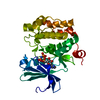
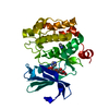
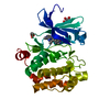
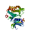
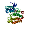
| Title | Crystal structure of PIM1 in complex with a Pyrrolo(1,2-a)Pyrazinone inhibitor | ||||||
 Components Components | SERINE/THREONINE-PROTEIN KINASE PIM-1 | ||||||
 Keywords Keywords |  TRANSFERASE / PIM1 / ATP BINDING / KINASE INHIBITOR TRANSFERASE / PIM1 / ATP BINDING / KINASE INHIBITOR | ||||||
| Function / homology |  Function and homology information Function and homology informationpositive regulation of cardioblast proliferation / cellular detoxification / regulation of hematopoietic stem cell proliferation / vitamin D receptor signaling pathway / STAT5 activation downstream of FLT3 ITD mutants / ribosomal small subunit binding / transcription factor binding / positive regulation of cyclin-dependent protein serine/threonine kinase activity / positive regulation of cardiac muscle cell proliferation / positive regulation of TORC1 signaling ...positive regulation of cardioblast proliferation / cellular detoxification / regulation of hematopoietic stem cell proliferation / vitamin D receptor signaling pathway / STAT5 activation downstream of FLT3 ITD mutants / ribosomal small subunit binding / transcription factor binding / positive regulation of cyclin-dependent protein serine/threonine kinase activity / positive regulation of cardiac muscle cell proliferation / positive regulation of TORC1 signaling / Signaling by FLT3 fusion proteins / positive regulation of brown fat cell differentiation / negative regulation of innate immune response / protein serine/threonine kinase activator activity / regulation of transmembrane transporter activity / positive regulation of protein serine/threonine kinase activity / negative regulation of DNA-binding transcription factor activity / cellular response to type II interferon / manganese ion binding / Interleukin-4 and Interleukin-13 signaling / protein autophosphorylation / protein stabilization / non-specific serine/threonine protein kinase / cell cycle / protein phosphorylation / protein serine kinase activity / protein serine/threonine kinase activity / apoptotic process / nucleolus / negative regulation of apoptotic process / positive regulation of DNA-templated transcription / nucleoplasm / ATP binding / nucleus / plasma membrane / cytosol / cytoplasmSimilarity search - Function | ||||||
| Biological species |  HOMO SAPIENS (human) HOMO SAPIENS (human) | ||||||
| Method | X-RAY DIFFRACTION / SYNCHROTRON / MOLECULAR REPLACEMENT / Resolution: 1.9 Å | ||||||
 Authors Authors | Casale, E. / Casuscelli, F. / Ardini, E. / Avanzi, N. / Cervi, G. / D'Anello, M. / Donati, D. / Faiardi, D. / Ferguson, R.D. / Fogliatto, G. ...Casale, E. / Casuscelli, F. / Ardini, E. / Avanzi, N. / Cervi, G. / D'Anello, M. / Donati, D. / Faiardi, D. / Ferguson, R.D. / Fogliatto, G. / Galvani, A. / Marsiglio, A. / Mirizzi, D.G. / Montemartini, M. / Orrenius, C. / Papeo, G. / Piutti, C. / Salom, B. / Felder, E.R. | ||||||
 Citation Citation | Journal: Bioorg.Med.Chem. / Year: 2013 Title: Discovery and Optimization of Pyrrolo[1,2-A]Pyrazinones Leads to Novel and Selective Inhibitors of Pim Kinases. Authors: Casuscelli, F. / Ardini, E. / Avanzi, N. / Casale, E. / Cervi, G. / D'Anello, M. / Donati, D. / Faiardi, D. / Ferguson, R.D. / Fogliatto, G. / Galvani, A. / Marsiglio, A. / Mirizzi, D.G. / ...Authors: Casuscelli, F. / Ardini, E. / Avanzi, N. / Casale, E. / Cervi, G. / D'Anello, M. / Donati, D. / Faiardi, D. / Ferguson, R.D. / Fogliatto, G. / Galvani, A. / Marsiglio, A. / Mirizzi, D.G. / Montemartini, M. / Orrenius, C. / Papeo, G. / Piutti, C. / Salom, B. / Felder, E.R. | ||||||
| History |
|
- Structure visualization
| Structure viewer | Molecule: MolmilJmol/JSmol |
|---|
- Downloads & links
-Download
| PDBx/mmCIF format | 4bzn.cif.gz | 75.1 KB | Display | PDBx/mmCIF format |
|---|---|---|---|---|
| PDB format | pdb4bzn.ent.gz | 54.4 KB | Display | PDB format |
| PDBx/mmJSON format | 4bzn.json.gz | Tree view | PDBx/mmJSON format | |
| Others |  Other downloads Other downloads |
-Validation report
| Arichive directory | https://data.pdbj.org/pub/pdb/validation_reports/bz/4bznftp://data.pdbj.org/pub/pdb/validation_reports/bz/4bzn | HTTPS FTP |
|---|
-Related structure data
| Related structure data | 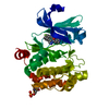 4bzoC  1yxtS C: citing same article ( S: Starting model for refinement |
|---|---|
| Similar structure data |


-Links
 PDBj
PDBj

- Assembly
| Deposited unit | 
| ||||||||
|---|---|---|---|---|---|---|---|---|---|
| 1 |
| ||||||||
| Unit cell |
|
-Components
| #1: Protein | Mass: 35835.691 Da / Num. of mol.: 1 / Fragment: KINASE DOMAIN, RESIDUES 2-313 Source method: isolated from a genetically manipulated source Source: (gene. exp.) HOMO SAPIENS (human) / Production host:  ESCHERICHIA COLI (E. coli) ESCHERICHIA COLI (E. coli)References: UniProt: P11309, non-specific serine/threonine protein kinase |
|---|---|
| #2: Chemical | ChemComp-UGX /   Mass: 345.459 Da / Num. of mol.: 1 / Source method: obtained synthetically / Formula: C18H23N3O2S Mass: 345.459 Da / Num. of mol.: 1 / Source method: obtained synthetically / Formula: C18H23N3O2S |
| #3: Water | ChemComp-HOH / Water Mass: 18.015 Da / Num. of mol.: 170 / Source method: isolated from a natural source / Formula: H2O Mass: 18.015 Da / Num. of mol.: 170 / Source method: isolated from a natural source / Formula: H2O |
| Sequence details | THE FIRST TWO RESIDUES AT THE N-TERMINAL GLY AND PRO ARE EXPRESSION |
-Experimental details
-Experiment
| Experiment | Method: X-RAY DIFFRACTION / Number of used crystals: 1 |
|---|
- Sample preparation
| Crystal | Density Matthews: 3.19 Å3/Da / Density % sol: 61.15 % / Description: NONE |
|---|---|
| Crystal grow | Details: 20% PEG 3350K, 0.3 M NACL, 0.1 M TRISHCL PH 7.6 |
-Data collection
| Diffraction | Mean temperature: 100 K |
|---|---|
| Diffraction source | Source: SYNCHROTRON / Site: ESRF  / Beamline: ID14-4 / Wavelength: 0.97 / Beamline: ID14-4 / Wavelength: 0.97 |
| Detector | Type: MARRESEARCH / Detector: CCD / Date: Apr 29, 2008 |
| Radiation | Protocol: SINGLE WAVELENGTH / Monochromatic (M) / Laue (L): M / Scattering type: x-ray |
| Radiation wavelength | Wavelength: 0.97 Å / Relative weight: 1 |
| Reflection | Resolution: 1.9→30 Å / Num. obs: 33517 / % possible obs: 99.7 % / Observed criterion σ(I): 0 / Redundancy: 3.6 % / Rmerge(I) obs: 0.06 / Net I/σ(I): 12.3 |
| Reflection shell | Resolution: 1.9→1.97 Å / Rmerge(I) obs: 0.47 / Mean I/σ(I) obs: 2.7 / % possible all: 99.6 |
- Processing
| Software |
| ||||||||||||||||||||||||||||||||||||||||||||||||||||||||||||||||||||||||||||||||||||||||||||||||||||||||||||||||||||||||||||||||||||||||||||||||||||||||||||||||||||||||||||||||||||||
|---|---|---|---|---|---|---|---|---|---|---|---|---|---|---|---|---|---|---|---|---|---|---|---|---|---|---|---|---|---|---|---|---|---|---|---|---|---|---|---|---|---|---|---|---|---|---|---|---|---|---|---|---|---|---|---|---|---|---|---|---|---|---|---|---|---|---|---|---|---|---|---|---|---|---|---|---|---|---|---|---|---|---|---|---|---|---|---|---|---|---|---|---|---|---|---|---|---|---|---|---|---|---|---|---|---|---|---|---|---|---|---|---|---|---|---|---|---|---|---|---|---|---|---|---|---|---|---|---|---|---|---|---|---|---|---|---|---|---|---|---|---|---|---|---|---|---|---|---|---|---|---|---|---|---|---|---|---|---|---|---|---|---|---|---|---|---|---|---|---|---|---|---|---|---|---|---|---|---|---|---|---|---|---|
| Refinement | Method to determine structure: MOLECULAR REPLACEMENT Starting model: PDB ENTRY 1YXT Resolution: 1.9→30 Å / Cor.coef. Fo:Fc: 0.966 / Cor.coef. Fo:Fc free: 0.958 / SU B: 2.341 / SU ML: 0.07 / Cross valid method: THROUGHOUT / ESU R: 0.113 / ESU R Free: 0.106 / Stereochemistry target values: MAXIMUM LIKELIHOOD / Details: HYDROGENS HAVE BEEN ADDED IN THE RIDING POSITIONS.
| ||||||||||||||||||||||||||||||||||||||||||||||||||||||||||||||||||||||||||||||||||||||||||||||||||||||||||||||||||||||||||||||||||||||||||||||||||||||||||||||||||||||||||||||||||||||
| Solvent computation | Ion probe radii: 0.8 Å / Shrinkage radii: 0.8 Å / VDW probe radii: 1.2 Å / Solvent model: MASK | ||||||||||||||||||||||||||||||||||||||||||||||||||||||||||||||||||||||||||||||||||||||||||||||||||||||||||||||||||||||||||||||||||||||||||||||||||||||||||||||||||||||||||||||||||||||
| Displacement parameters | Biso mean: 35.881 Å2
| ||||||||||||||||||||||||||||||||||||||||||||||||||||||||||||||||||||||||||||||||||||||||||||||||||||||||||||||||||||||||||||||||||||||||||||||||||||||||||||||||||||||||||||||||||||||
| Refinement step | Cycle: LAST / Resolution: 1.9→30 Å
| ||||||||||||||||||||||||||||||||||||||||||||||||||||||||||||||||||||||||||||||||||||||||||||||||||||||||||||||||||||||||||||||||||||||||||||||||||||||||||||||||||||||||||||||||||||||
| Refine LS restraints |
|