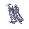
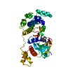
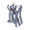
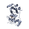
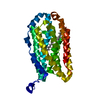
Mass: 35890.828 Da / Num. of mol.: 1 / Mutation: YES Source method: isolated from a genetically manipulated source Details: RESIDUES 3-32 AT THE N-TERMINUS AND RESIDUES 244-271 OF THE THIRD INTRACELLULAR LOOP WERE DELETED FROM THE CONSTRUCT. THE CONSTRUCT WAS TRUNCATED AFTER RESIDUE 367 AND A HEXAHIS TAG ADDED. Source: (gene. exp.) MELEAGRIS GALLOPAVO (turkey) / Cell: ERYTHROCYTE / Plasmid: PBACPAK8 / Cell line (production host): High Five / Production host: TRICHOPLUSIA NI (cabbage looper) / References: UniProt: P07700
Mass: 18.015 Da / Num. of mol.: 38 / Source method: isolated from a natural source / Formula: H2O
-
Details
Sequence details
THE FOLLOWING MUTATIONS WERE MADE TO IMPROVE THERMOSTABILITY ...THE FOLLOWING MUTATIONS WERE MADE TO IMPROVE THERMOSTABILITY R68S,M90V,I129V,Y227A,A282L,D322K,F327A, F338M,Y343L. THE FOLLOWING MUTATIONS WERE MADE TO IMPROVE EXPRESSION AND HELP CRYSTALLISATION C116L, C358A
-
Experimental details
-
Experiment
Experiment
Method: X-RAY DIFFRACTION / Number of used crystals: 1
-
Sample preparation
Crystal
Density Matthews: 2.19 Å3/Da / Density % sol: 44 % / Description: NONE
Crystal grow
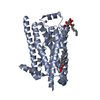
Temperature: 293 K / Method: lipidic cubic phase / pH: 7 Details: 25% PEG 600, 0.1M ADA PH7.0 LIPID CUBIC PHASE (LCP) TEMPERATURE 293K
Resolution: 2.1→30.88 Å / Cor.coef. Fo:Fc: 0.939 / Cor.coef. Fo:Fc free: 0.904 / SU B: 4.814 / SU ML: 0.131 / Cross valid method: THROUGHOUT / ESU R: 0.238 / ESU R Free: 0.199 / Stereochemistry target values: MAXIMUM LIKELIHOOD Details: HYDROGENS HAVE BEEN ADDED IN THE RIDING POSITIONS. U VALUES REFINED INDIVIDUALLY
Rfactor
Num. reflection
% reflection
Selection details
Rfree
0.24552
964
5.2 %
RANDOM
Rwork
0.19287
-
-
-
obs
0.19558
17696
97.5 %
-
Solvent computation
Ion probe radii: 0.8 Å / Shrinkage radii: 0.8 Å / VDW probe radii: 1.2 Å / Solvent model: MASK
 Movie
Movie Controller


 Open data
 Basic information
 Components
 Keywords
 Function and homology information
 Authors
 Citation
 Structure visualization
 Downloads & links
 Other downloads
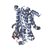


 PDBj
PDBj


 Assembly
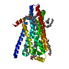


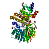
 Mass: 22.990 Da / Num. of mol.: 2 / Source method: obtained synthetically / Formula: Na
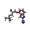
 Mass: 287.357 Da / Num. of mol.: 1 / Source method: obtained synthetically / Formula: C16H21N3O2
 Mass: 190.154 Da / Num. of mol.: 1 / Source method: obtained synthetically / Formula: C6H10N2O5 / Comment: pH buffer*YM
 Mass: 356.540 Da / Num. of mol.: 7 / Source method: obtained synthetically / Formula: C21H40O4
 Sample preparation
 / Beamline: ID23-2 / Wavelength: 0.8726
  Processing