 Movie
Movie Controller


+ Open data

- Basic information
| Entry | Database: PDB / ID: 4bk8 | ||||||
|---|---|---|---|---|---|---|---|
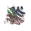
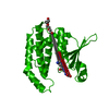
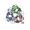
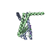
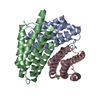
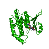
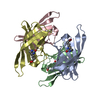
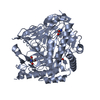
| Title | Superoxide reductase (Neelaredoxin) from Ignicoccus hospitalis | ||||||
 Components Components | DESULFOFERRODOXIN, FERROUS IRON-BINDING REGION | ||||||
 Keywords Keywords |  OXIDOREDUCTASE / NEELAREDOXIN / SYMBIOSIS / OXIDATIVE STRESS OXIDOREDUCTASE / NEELAREDOXIN / SYMBIOSIS / OXIDATIVE STRESS | ||||||
| Function / homology |  Function and homology information Function and homology information | ||||||
| Biological species |   IGNICOCCUS HOSPITALIS (archaea) IGNICOCCUS HOSPITALIS (archaea) | ||||||
| Method | X-RAY DIFFRACTION / SYNCHROTRON / MOLECULAR REPLACEMENT / Resolution: 1.847 Å | ||||||
 Authors Authors | Romao, C.V. / Matias, P.M. / Pinho, F.G. / Sousa, C.M. / Barradas, A.R. / Pinto, A.F. / Teixeira, M. / Bandeiras, T.M. | ||||||
 Citation Citation | Journal: To be Published Title: Structure of a Natural Sor Mutant Authors: Pinho, F.G. / Romao, C.V. / Pinto, A.F. / Matias, P.M. / Teixeira, M. / Bandeiras, T.M. #1: Journal: Acta Crystallogr.,Sect.F / Year: 2010 Title: Cloning, Purification, Crystallization and X-Ray Crystallographic Analysis of Ignicoccus Hospitalis Neelaredoxin. Authors: Pinho, F.G. / Romao, C.V. / Pinto, A.F. / Saraiva, L.M. / Huber, H. / Matias, P.M. / Teixeira, M. / Bandeiras, T.M. | ||||||
| History |
|
- Structure visualization
| Structure viewer | Molecule: MolmilJmol/JSmol |
|---|
- Downloads & links
-Download
| PDBx/mmCIF format | 4bk8.cif.gz | 61.9 KB | Display | PDBx/mmCIF format |
|---|---|---|---|---|
| PDB format | pdb4bk8.ent.gz | 50.2 KB | Display | PDB format |
| PDBx/mmJSON format | 4bk8.json.gz | Tree view | PDBx/mmJSON format | |
| Others |  Other downloads Other downloads |
-Validation report
| Arichive directory | https://data.pdbj.org/pub/pdb/validation_reports/bk/4bk8ftp://data.pdbj.org/pub/pdb/validation_reports/bk/4bk8 | HTTPS FTP |
|---|
-Related structure data


-Links
 PDBj
PDBj- Assembly
| Deposited unit | 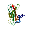
| ||||||||
|---|---|---|---|---|---|---|---|---|---|
| 1 | 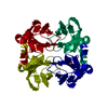
| ||||||||
| Unit cell |
|
-Components
| #1: Protein | Mass: 14113.092 Da / Num. of mol.: 1 Source method: isolated from a genetically manipulated source Source: (gene. exp.) IGNICOCCUS HOSPITALIS (archaea) / Production host:  ESCHERICHIA COLI (E. coli) / Strain (production host): BL21(DE3) / Variant (production host): GOLD / References: UniProt: A8AC72 ESCHERICHIA COLI (E. coli) / Strain (production host): BL21(DE3) / Variant (production host): GOLD / References: UniProt: A8AC72 |
|---|---|
| #2: Chemical | ChemComp-FE / Iron  Mass: 55.845 Da / Num. of mol.: 1 / Source method: obtained synthetically / Formula: Fe Mass: 55.845 Da / Num. of mol.: 1 / Source method: obtained synthetically / Formula: Fe |
| #3: Water | ChemComp-HOH / Water Mass: 18.015 Da / Num. of mol.: 102 / Source method: isolated from a natural source / Formula: H2O Mass: 18.015 Da / Num. of mol.: 102 / Source method: isolated from a natural source / Formula: H2O |
-Experimental details
-Experiment
| Experiment | Method: X-RAY DIFFRACTION / Number of used crystals: 1 |
|---|
- Sample preparation
| Crystal | Density Matthews: 3.76 Å3/Da / Density % sol: 67.32 % Description: THE 3D STRUCTURE WAS SOLVED FROM A SAD DATASET MEASURED IN-HOUSE TO 2.4A AND A PRELIMINARY REFINEMENT CARRIED OUT. FOLLOWING A MOLECULAR REPLACEMENT STEP TO CORRECTLY POSITION THE ...Description: THE 3D STRUCTURE WAS SOLVED FROM A SAD DATASET MEASURED IN-HOUSE TO 2.4A AND A PRELIMINARY REFINEMENT CARRIED OUT. FOLLOWING A MOLECULAR REPLACEMENT STEP TO CORRECTLY POSITION THE RESULTING MODEL IN THE UNIT CELL OF THE CRYSTAL MEASURED AT THE ESRF, IT WAS USED IN THE FINAL REFINEMENT. |
|---|---|
| Crystal grow | pH: 8.5 Details: 85 MM TRISHCL PH 8.5, 8.5 MM NICL2, 15%(V/V) PEG 2000 MONOMETHYL ETHER AND 15%(V/V) GLYCEROL |
-Data collection
| Diffraction | Mean temperature: 100 K |
|---|---|
| Diffraction source | Source: SYNCHROTRON / Site: ESRF  / Beamline: ID23-1 / Wavelength: 0.98 / Beamline: ID23-1 / Wavelength: 0.98 |
| Detector | Type: ADSC QUANTUM 315r / Detector: CCD / Date: Jul 14, 2009 / Details: BENT CYLINDRICAL MIRROR |
| Radiation | Monochromator: SI(111) / Protocol: SINGLE WAVELENGTH / Monochromatic (M) / Laue (L): M / Scattering type: x-ray |
| Radiation wavelength | Wavelength: 0.98 Å / Relative weight: 1 |
| Reflection | Resolution: 1.85→47.2 Å / Num. obs: 18718 / % possible obs: 98.6 % / Observed criterion σ(I): 0 / Redundancy: 11.6 % / Rmerge(I) obs: 0.1 / Net I/σ(I): 18.23 |
| Reflection shell | Resolution: 1.85→1.96 Å / Redundancy: 11.6 % / Rmerge(I) obs: 0.92 / Mean I/σ(I) obs: 2.14 / % possible all: 98 |
- Processing
| Software |
| ||||||||||||||||||||||||||||||||||||||||||||||||||||||||||||||||||||||||||||||||||||||||||||||||||||||||||||||||||||||||||||||||||||||||||||||||||||||||||||||||||||||||||||||||||||||||||||||||||||||||||||||||||||||||||||||||||||||||||||||||||||||||||
|---|---|---|---|---|---|---|---|---|---|---|---|---|---|---|---|---|---|---|---|---|---|---|---|---|---|---|---|---|---|---|---|---|---|---|---|---|---|---|---|---|---|---|---|---|---|---|---|---|---|---|---|---|---|---|---|---|---|---|---|---|---|---|---|---|---|---|---|---|---|---|---|---|---|---|---|---|---|---|---|---|---|---|---|---|---|---|---|---|---|---|---|---|---|---|---|---|---|---|---|---|---|---|---|---|---|---|---|---|---|---|---|---|---|---|---|---|---|---|---|---|---|---|---|---|---|---|---|---|---|---|---|---|---|---|---|---|---|---|---|---|---|---|---|---|---|---|---|---|---|---|---|---|---|---|---|---|---|---|---|---|---|---|---|---|---|---|---|---|---|---|---|---|---|---|---|---|---|---|---|---|---|---|---|---|---|---|---|---|---|---|---|---|---|---|---|---|---|---|---|---|---|---|---|---|---|---|---|---|---|---|---|---|---|---|---|---|---|---|---|---|---|---|---|---|---|---|---|---|---|---|---|---|---|---|---|---|---|---|---|---|---|---|---|---|---|---|---|---|---|---|---|
| Refinement | Method to determine structure: MOLECULAR REPLACEMENT Starting model: PRELIMINARY MODEL FROM SAD Resolution: 1.847→47.196 Å / SU ML: 0.14 / σ(F): 1.88 / Phase error: 22.61 / Stereochemistry target values: ML
| ||||||||||||||||||||||||||||||||||||||||||||||||||||||||||||||||||||||||||||||||||||||||||||||||||||||||||||||||||||||||||||||||||||||||||||||||||||||||||||||||||||||||||||||||||||||||||||||||||||||||||||||||||||||||||||||||||||||||||||||||||||||||||
| Solvent computation | Shrinkage radii: 0.9 Å / VDW probe radii: 1.11 Å / Solvent model: FLAT BULK SOLVENT MODEL | ||||||||||||||||||||||||||||||||||||||||||||||||||||||||||||||||||||||||||||||||||||||||||||||||||||||||||||||||||||||||||||||||||||||||||||||||||||||||||||||||||||||||||||||||||||||||||||||||||||||||||||||||||||||||||||||||||||||||||||||||||||||||||
| Refinement step | Cycle: LAST / Resolution: 1.847→47.196 Å
| ||||||||||||||||||||||||||||||||||||||||||||||||||||||||||||||||||||||||||||||||||||||||||||||||||||||||||||||||||||||||||||||||||||||||||||||||||||||||||||||||||||||||||||||||||||||||||||||||||||||||||||||||||||||||||||||||||||||||||||||||||||||||||
| Refine LS restraints |
| ||||||||||||||||||||||||||||||||||||||||||||||||||||||||||||||||||||||||||||||||||||||||||||||||||||||||||||||||||||||||||||||||||||||||||||||||||||||||||||||||||||||||||||||||||||||||||||||||||||||||||||||||||||||||||||||||||||||||||||||||||||||||||
| LS refinement shell |
| ||||||||||||||||||||||||||||||||||||||||||||||||||||||||||||||||||||||||||||||||||||||||||||||||||||||||||||||||||||||||||||||||||||||||||||||||||||||||||||||||||||||||||||||||||||||||||||||||||||||||||||||||||||||||||||||||||||||||||||||||||||||||||
| Refinement TLS params. | Method: refined / Refine-ID: X-RAY DIFFRACTION
| ||||||||||||||||||||||||||||||||||||||||||||||||||||||||||||||||||||||||||||||||||||||||||||||||||||||||||||||||||||||||||||||||||||||||||||||||||||||||||||||||||||||||||||||||||||||||||||||||||||||||||||||||||||||||||||||||||||||||||||||||||||||||||
| Refinement TLS group |
|
