 Movie
Movie Controller

[English] 日本語
 Yorodumi
- PDB-4bet: Crystal structure of the Legionella pneumophila FIC domain-contai... -
+ Open data

- Basic information
| Entry | Database: PDB / ID: 4bet | ||||||
|---|---|---|---|---|---|---|---|
| Title | Crystal structure of the Legionella pneumophila FIC domain-containing effector AnkX protein (inactive H229A mutant) in complex with cytidine-diphosphate-choline | ||||||
 Components Components | PHOSPHOCHOLINE TRANSFERASE ANKX | ||||||
 Keywords Keywords |  TRANSFERASE / PHOSPHOCHOLINATION / TYPE IV SECRETION SYSTEM EFFECTOR / CYTIDINE- DIPHOSPHATE-CHOLINE TRANSFERASE / PHOSPHOCHOLINATION / TYPE IV SECRETION SYSTEM EFFECTOR / CYTIDINE- DIPHOSPHATE-CHOLINE | ||||||
| Function / homology |  Function and homology information Function and homology informationphosphocholine transferase activity / Transferases; Transferring phosphorus-containing groups; Phosphotransferases with an alcohol group as acceptor / regulation of GTPase activity / host cell cytoplasm / extracellular regionSimilarity search - Function | ||||||
| Biological species |   LEGIONELLA PNEUMOPHILA (bacteria) LEGIONELLA PNEUMOPHILA (bacteria) | ||||||
| Method | X-RAY DIFFRACTION / SYNCHROTRON / MOLECULAR REPLACEMENT / Resolution: 2.55 Å | ||||||
 Authors Authors | Campanacci, V. / Mukherjee, S. / Roy, C.R. / Cherfils, J. | ||||||
 Citation Citation | Journal: Embo J. / Year: 2013 Title: Structure of the Legionella Effector Ankx Reveals the Mechanism of Phosphocholine Transfer by the Fic Domain. Authors: Campanacci, V. / Mukherjee, S. / Roy, C.R. / Cherfils, J. | ||||||
| History |
|
- Structure visualization
| Structure viewer | Molecule: MolmilJmol/JSmol |
|---|
- Downloads & links
-Download
| PDBx/mmCIF format | 4bet.cif.gz | 399.2 KB | Display | PDBx/mmCIF format |
|---|---|---|---|---|
| PDB format | pdb4bet.ent.gz | 331.6 KB | Display | PDB format |
| PDBx/mmJSON format | 4bet.json.gz | Tree view | PDBx/mmJSON format | |
| Others |  Other downloads Other downloads |
-Validation report
| Arichive directory | https://data.pdbj.org/pub/pdb/validation_reports/be/4betftp://data.pdbj.org/pub/pdb/validation_reports/be/4bet | HTTPS FTP |
|---|
-Related structure data
| Related structure data |  4bepSC  4berC  4besC S: Starting model for refinement C: citing same article ( |
|---|---|
| Similar structure data |


-Links
 PDBj
PDBj- Assembly
| Deposited unit | 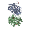
| ||||||||
|---|---|---|---|---|---|---|---|---|---|
| 1 | 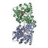
| ||||||||
| 2 | 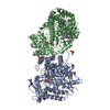
| ||||||||
| Unit cell |
| ||||||||
| Noncrystallographic symmetry (NCS) | NCS oper: (Code: given Matrix: (-0.470724, 0.003868, 0.882272), Vector : |
-Components
| #1: Protein | Mass: 58284.461 Da / Num. of mol.: 2 / Fragment: FIC AND ANKYRIN REPEATS DOMAINS, RESIDUES 2-484 / Mutation: YES Source method: isolated from a genetically manipulated source Details: INACTIVE MUTANT / Source: (gene. exp.) LEGIONELLA PNEUMOPHILA (bacteria) / Strain: PHILADELPHIA 1 / Production host: ESCHERICHIA COLI (E. coli) / Strain (production host): BL21(DE3) / Variant (production host): ROSETTA PLYSSReferences: UniProt: Q5ZXN6, Transferases; Transferring phosphorus-containing groups; Phosphotransferases with an alcohol group as acceptor#2: Chemical | Citicoline  Mass: 488.324 Da / Num. of mol.: 2 / Source method: obtained synthetically / Formula: C14H26N4O11P2 Mass: 488.324 Da / Num. of mol.: 2 / Source method: obtained synthetically / Formula: C14H26N4O11P2#3: Chemical | ChemComp-SO4 / Sulfate  Mass: 96.063 Da / Num. of mol.: 4 / Source method: obtained synthetically / Formula: SO4 Mass: 96.063 Da / Num. of mol.: 4 / Source method: obtained synthetically / Formula: SO4#4: Chemical | Glycerol  Mass: 92.094 Da / Num. of mol.: 3 / Source method: obtained synthetically / Formula: C3H8O3 Mass: 92.094 Da / Num. of mol.: 3 / Source method: obtained synthetically / Formula: C3H8O3#5: Water | ChemComp-HOH / | Water Mass: 18.015 Da / Num. of mol.: 114 / Source method: isolated from a natural source / Formula: H2O Mass: 18.015 Da / Num. of mol.: 114 / Source method: isolated from a natural source / Formula: H2O |
|---|
-Experimental details
-Experiment
| Experiment | Method: X-RAY DIFFRACTION / Number of used crystals: 1 |
|---|
- Sample preparation
| Crystal | Density Matthews: 2.5 Å3/Da / Density % sol: 50.9 % / Description: NONE |
|---|---|
| Crystal grow | pH: 7.5 Details: 0.2 M LITHIUM SULFATE, 14% PEG 4000, 0.1 M TRIS PH 7.5 |
-Data collection
| Diffraction | Mean temperature: 100 K |
|---|---|
| Diffraction source | Source: SYNCHROTRON / Site: SOLEIL  / Beamline: PROXIMA 1 / Wavelength: 0.9801 / Beamline: PROXIMA 1 / Wavelength: 0.9801 |
| Detector | Type: ADSC QUANTUM 315r / Detector: CCD / Date: May 2, 2012 / Details: MIRRORS |
| Radiation | Protocol: SINGLE WAVELENGTH / Monochromatic (M) / Laue (L): M / Scattering type: x-ray |
| Radiation wavelength | Wavelength: 0.9801 Å / Relative weight: 1 |
| Reflection | Resolution: 2.55→43.89 Å / Num. obs: 36935 / % possible obs: 98.7 % / Redundancy: 4.7 % / Biso Wilson estimate: 60.66 Å2 / Rmerge(I) obs: 0.09 / Net I/σ(I): 12.02 |
| Reflection shell | Resolution: 2.55→2.7 Å / Redundancy: 4.7 % / Rmerge(I) obs: 0.84 / Mean I/σ(I) obs: 1.75 / % possible all: 97.4 |
- Processing
| Software |
| |||||||||||||||||||||||||||||||||||||||||||||||||||||||||||||||||||||||||||||||||||||||||||||||||||||||||||||||||||||||||||||||||||||||||||||||||||||||||||||||||||||||||||||||
|---|---|---|---|---|---|---|---|---|---|---|---|---|---|---|---|---|---|---|---|---|---|---|---|---|---|---|---|---|---|---|---|---|---|---|---|---|---|---|---|---|---|---|---|---|---|---|---|---|---|---|---|---|---|---|---|---|---|---|---|---|---|---|---|---|---|---|---|---|---|---|---|---|---|---|---|---|---|---|---|---|---|---|---|---|---|---|---|---|---|---|---|---|---|---|---|---|---|---|---|---|---|---|---|---|---|---|---|---|---|---|---|---|---|---|---|---|---|---|---|---|---|---|---|---|---|---|---|---|---|---|---|---|---|---|---|---|---|---|---|---|---|---|---|---|---|---|---|---|---|---|---|---|---|---|---|---|---|---|---|---|---|---|---|---|---|---|---|---|---|---|---|---|---|---|---|---|
| Refinement | Method to determine structure: MOLECULAR REPLACEMENT Starting model: PDB ENTRY 4BEP Resolution: 2.55→43.89 Å / Cor.coef. Fo:Fc: 0.9232 / Cor.coef. Fo:Fc free: 0.8891 / SU R Cruickshank DPI: 0.637 / Cross valid method: THROUGHOUT / σ(F): 0 / SU R Blow DPI: 0.545 / SU Rfree Blow DPI: 0.281 / SU Rfree Cruickshank DPI: 0.291 Details: IDEAL-DIST CONTACT TERM CONTACT SETUP. ALL ATOMS HAVE CCP4 ATOM TYPE FROM LIBRARY
| |||||||||||||||||||||||||||||||||||||||||||||||||||||||||||||||||||||||||||||||||||||||||||||||||||||||||||||||||||||||||||||||||||||||||||||||||||||||||||||||||||||||||||||||
| Displacement parameters | Biso mean: 69.76 Å2
| |||||||||||||||||||||||||||||||||||||||||||||||||||||||||||||||||||||||||||||||||||||||||||||||||||||||||||||||||||||||||||||||||||||||||||||||||||||||||||||||||||||||||||||||
| Refine analyze | Luzzati coordinate error obs: 0.4 Å | |||||||||||||||||||||||||||||||||||||||||||||||||||||||||||||||||||||||||||||||||||||||||||||||||||||||||||||||||||||||||||||||||||||||||||||||||||||||||||||||||||||||||||||||
| Refinement step | Cycle: LAST / Resolution: 2.55→43.89 Å
| |||||||||||||||||||||||||||||||||||||||||||||||||||||||||||||||||||||||||||||||||||||||||||||||||||||||||||||||||||||||||||||||||||||||||||||||||||||||||||||||||||||||||||||||
| Refine LS restraints |
| |||||||||||||||||||||||||||||||||||||||||||||||||||||||||||||||||||||||||||||||||||||||||||||||||||||||||||||||||||||||||||||||||||||||||||||||||||||||||||||||||||||||||||||||
| LS refinement shell | Resolution: 2.55→2.62 Å / Total num. of bins used: 19
| |||||||||||||||||||||||||||||||||||||||||||||||||||||||||||||||||||||||||||||||||||||||||||||||||||||||||||||||||||||||||||||||||||||||||||||||||||||||||||||||||||||||||||||||
| Refinement TLS params. | Method: refined / Refine-ID: X-RAY DIFFRACTION
| |||||||||||||||||||||||||||||||||||||||||||||||||||||||||||||||||||||||||||||||||||||||||||||||||||||||||||||||||||||||||||||||||||||||||||||||||||||||||||||||||||||||||||||||
| Refinement TLS group |
|
