 Movie
Movie Controller

[English] 日本語
 Yorodumi
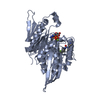
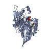
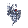
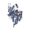
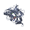
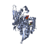
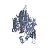
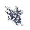
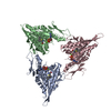
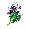
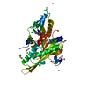
- PDB-4bbg: Crystal structure of human kinesin Eg5 in complex with 3-(((2-Ami... -
+ Open data

- Basic information
| Entry | Database: PDB / ID: 4bbg | ||||||
|---|---|---|---|---|---|---|---|
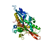
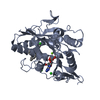
| Title | Crystal structure of human kinesin Eg5 in complex with 3-(((2-Aminoethyl)sulfanyl)(3-ethylphenyl) phenylmethyl)phenol | ||||||
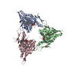
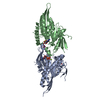
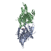
 Components Components | KINESIN-LIKE PROTEIN KIF11 | ||||||
 Keywords Keywords | CELL CYCLE / MITOSIS / INHIBITOR | ||||||
| Function / homology |  Function and homology information Function and homology informationspindle elongation / Kinesins / plus-end-directed microtubule motor activity / regulation of mitotic centrosome separation / mitotic centrosome separation / COPI-dependent Golgi-to-ER retrograde traffic / kinesin complex / microtubule motor activity / spindle organization / microtubule-based movement ...spindle elongation / Kinesins / plus-end-directed microtubule motor activity / regulation of mitotic centrosome separation / mitotic centrosome separation / COPI-dependent Golgi-to-ER retrograde traffic / kinesin complex / microtubule motor activity / spindle organization / microtubule-based movement / mitotic spindle assembly / MHC class II antigen presentation / mitotic spindle organization / mitotic spindle / spindle / spindle pole / mitotic cell cycle / microtubule binding / microtubule / cell division / protein kinase binding / protein-containing complex / ATP binding / membrane / nucleus / cytosolSimilarity search - Function | ||||||
| Biological species |  HOMO SAPIENS (human) HOMO SAPIENS (human) | ||||||
| Method | X-RAY DIFFRACTION / SYNCHROTRON / MOLECULAR REPLACEMENT / Resolution: 2.75 Å | ||||||
 Authors Authors | Kaan, H.Y.K. / Kozielski, F. | ||||||
 Citation Citation | Journal: J.Med.Chem. / Year: 2013 Title: Optimized S-Trityl-L-Cysteine-Based Inhibitors of Kinesin Spindle Protein with Potent in Vivo Antitumor Activity in Lung Cancer Xenograft Models. Authors: Good, J.A.D. / Wang, F. / Rath, O. / Kaan, H.Y.K. / Talapatra, S.K. / Podgorski, D. / Mackay, S.P. / Kozielski, F. | ||||||
| History |
|
- Structure visualization
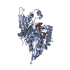
| Structure viewer | Molecule: MolmilJmol/JSmol |
|---|
- Downloads & links
-Download
| PDBx/mmCIF format | 4bbg.cif.gz | 86.5 KB | Display | PDBx/mmCIF format |
|---|---|---|---|---|
| PDB format | pdb4bbg.ent.gz | 64 KB | Display | PDB format |
| PDBx/mmJSON format | 4bbg.json.gz | Tree view | PDBx/mmJSON format | |
| Others |  Other downloads Other downloads |
-Validation report
| Arichive directory | https://data.pdbj.org/pub/pdb/validation_reports/bb/4bbgftp://data.pdbj.org/pub/pdb/validation_reports/bb/4bbg | HTTPS FTP |
|---|
-Related structure data
| Related structure data | 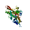 3kenS S: Starting model for refinement |
|---|---|
| Similar structure data |


-Links
 PDBj
PDBj


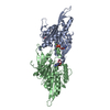
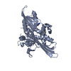
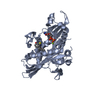
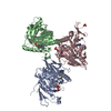
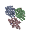
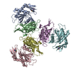
- Assembly
| Deposited unit | 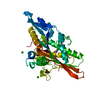
| ||||||||
|---|---|---|---|---|---|---|---|---|---|
| 1 | 
| ||||||||
| Unit cell |
|
-Components
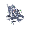
-Protein , 1 types, 1 molecules A
| #1: Protein | / KINESIN-LIKE PROTEIN 1 / KINESIN-LIKE SPINDLE PROTEIN HKSP / KINESIN-RELATED MOTOR PROTEIN EG5 / ...KINESIN-LIKE PROTEIN 1 / KINESIN-LIKE SPINDLE PROTEIN HKSP / KINESIN-RELATED MOTOR PROTEIN EG5 / THYROID RECEPTOR-INTERACTING PROTEIN 5 / TR-INTERACTING PROTEIN 5 / TRIP-5 Mass: 41055.582 Da / Num. of mol.: 1 / Fragment: MOTOR DOMAIN, RESIDUES 1 - 368 Source method: isolated from a genetically manipulated source Source: (gene. exp.) HOMO SAPIENS (human) / Production host:  ESCHERICHIA COLI (E. coli) / Strain (production host): BL21(DE3) / Variant (production host): PLYSS / References: UniProt: P52732 ESCHERICHIA COLI (E. coli) / Strain (production host): BL21(DE3) / Variant (production host): PLYSS / References: UniProt: P52732 |
|---|
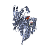
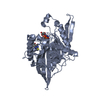
-Non-polymers , 5 types, 101 molecules 


| #2: Chemical | ChemComp-ADP / Adenosine diphosphate Mass: 427.201 Da / Num. of mol.: 1 / Source method: obtained synthetically / Formula: C10H15N5O10P2 / Comment: ADP, energy-carrying molecule*YM Mass: 427.201 Da / Num. of mol.: 1 / Source method: obtained synthetically / Formula: C10H15N5O10P2 / Comment: ADP, energy-carrying molecule*YM | ||||
|---|---|---|---|---|---|
| #3: Chemical | ChemComp-MG /  Mass: 24.305 Da / Num. of mol.: 1 / Source method: obtained synthetically / Formula: Mg Mass: 24.305 Da / Num. of mol.: 1 / Source method: obtained synthetically / Formula: Mg | ||||
| #4: Chemical | ChemComp-CL / Chloride Mass: 35.453 Da / Num. of mol.: 5 / Source method: obtained synthetically / Formula: Cl Mass: 35.453 Da / Num. of mol.: 5 / Source method: obtained synthetically / Formula: Cl#5: Chemical | ChemComp-V02 / |  Mass: 363.516 Da / Num. of mol.: 1 / Source method: obtained synthetically / Formula: C23H25NOS Mass: 363.516 Da / Num. of mol.: 1 / Source method: obtained synthetically / Formula: C23H25NOS#6: Water | ChemComp-HOH / | WaterMass: 18.015 Da / Num. of mol.: 93 / Source method: isolated from a natural source / Formula: H2O |
-Experimental details
-Experiment
| Experiment | Method: X-RAY DIFFRACTION / Number of used crystals: 1 |
|---|
- Sample preparation
| Crystal | Density Matthews: 4.02 Å3/Da / Density % sol: 69.44 % / Description: NONE |
|---|---|
| Crystal grow | pH: 5.5 Details: 22 % POLYETHYLENE GLYCOL-3350, 0.3 M AMMONIUM SULPHATE, 0.1 M MES PH 5.5 AND 0.01 M TRIMETHYLAMINE HYDROCHLORIDE |
-Data collection
| Diffraction | Mean temperature: 93 K |
|---|---|
| Diffraction source | Source: SYNCHROTRON / Site: ESRF  / Beamline: ID23-2 / Wavelength: 0.873 / Beamline: ID23-2 / Wavelength: 0.873 |
| Detector | Type: MARMOSAIC 225 mm CCD / Detector: CCD / Date: Nov 18, 2010 |
| Radiation | Protocol: SINGLE WAVELENGTH / Monochromatic (M) / Laue (L): M / Scattering type: x-ray |
| Radiation wavelength | Wavelength: 0.873 Å / Relative weight: 1 |
| Reflection | Resolution: 2.75→30 Å / Num. obs: 17253 / % possible obs: 99.9 % / Observed criterion σ(I): 2 / Redundancy: 7.6 % / Rmerge(I) obs: 0.08 / Net I/σ(I): 17 |
| Reflection shell | Resolution: 2.75→2.9 Å / Redundancy: 7.6 % / Rmerge(I) obs: 0.43 / Mean I/σ(I) obs: 5.3 / % possible all: 100 |
- Processing
| Software |
| ||||||||||||||||||||||||||||||||||||||||||||||||||||||||||||||||||||||||||||||||||||||||||||||||||||||||||||||||||||||||||||||||||||||||||||||||||||||||||||||||||||||||||||||||||||||
|---|---|---|---|---|---|---|---|---|---|---|---|---|---|---|---|---|---|---|---|---|---|---|---|---|---|---|---|---|---|---|---|---|---|---|---|---|---|---|---|---|---|---|---|---|---|---|---|---|---|---|---|---|---|---|---|---|---|---|---|---|---|---|---|---|---|---|---|---|---|---|---|---|---|---|---|---|---|---|---|---|---|---|---|---|---|---|---|---|---|---|---|---|---|---|---|---|---|---|---|---|---|---|---|---|---|---|---|---|---|---|---|---|---|---|---|---|---|---|---|---|---|---|---|---|---|---|---|---|---|---|---|---|---|---|---|---|---|---|---|---|---|---|---|---|---|---|---|---|---|---|---|---|---|---|---|---|---|---|---|---|---|---|---|---|---|---|---|---|---|---|---|---|---|---|---|---|---|---|---|---|---|---|---|
| Refinement | Method to determine structure: MOLECULAR REPLACEMENT Starting model: PDB ENTRY 3KEN Resolution: 2.75→30 Å / Cor.coef. Fo:Fc: 0.949 / Cor.coef. Fo:Fc free: 0.916 / SU B: 8.799 / SU ML: 0.183 / Cross valid method: THROUGHOUT / ESU R: 0.372 / ESU R Free: 0.265 / Stereochemistry target values: MAXIMUM LIKELIHOOD Details: HYDROGENS HAVE BEEN ADDED IN THE RIDING POSITIONS. U VALUES REFINED INDIVIDUALLY
| ||||||||||||||||||||||||||||||||||||||||||||||||||||||||||||||||||||||||||||||||||||||||||||||||||||||||||||||||||||||||||||||||||||||||||||||||||||||||||||||||||||||||||||||||||||||
| Solvent computation | Ion probe radii: 0.8 Å / Shrinkage radii: 0.8 Å / VDW probe radii: 1.4 Å / Solvent model: MASK | ||||||||||||||||||||||||||||||||||||||||||||||||||||||||||||||||||||||||||||||||||||||||||||||||||||||||||||||||||||||||||||||||||||||||||||||||||||||||||||||||||||||||||||||||||||||
| Displacement parameters | Biso mean: 55.846 Å2 | ||||||||||||||||||||||||||||||||||||||||||||||||||||||||||||||||||||||||||||||||||||||||||||||||||||||||||||||||||||||||||||||||||||||||||||||||||||||||||||||||||||||||||||||||||||||
| Refinement step | Cycle: LAST / Resolution: 2.75→30 Å
| ||||||||||||||||||||||||||||||||||||||||||||||||||||||||||||||||||||||||||||||||||||||||||||||||||||||||||||||||||||||||||||||||||||||||||||||||||||||||||||||||||||||||||||||||||||||
| Refine LS restraints |
|