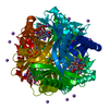
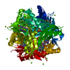
 Movie
Movie Controller


+ Open data

- Basic information
| Entry | Database: PDB / ID: 4b2j | ||||||
|---|---|---|---|---|---|---|---|
| Title | COMPLEXES OF DODECIN WITH FLAVIN AND FLAVIN-LIKE LIGANDS | ||||||
 Components Components | DODECIN | ||||||
 Keywords Keywords |  FLAVOPROTEIN / INCORPORATION / INCORPORATION OF ARTIFICIAL (NON-CANONICAL) AMINO ACIDS / PROTEIN REACTION CONTROL FLAVOPROTEIN / INCORPORATION / INCORPORATION OF ARTIFICIAL (NON-CANONICAL) AMINO ACIDS / PROTEIN REACTION CONTROL | ||||||
| Function / homology |  Function and homology information Function and homology information | ||||||
| Biological species |  HALOBACTERIUM SALINARUM (Halophile) HALOBACTERIUM SALINARUM (Halophile) | ||||||
| Method | X-RAY DIFFRACTION / SYNCHROTRON / MOLECULAR REPLACEMENT / Resolution: 1.9 Å | ||||||
 Authors Authors | Staudt, H. / Hoesl, M. / Dreuw, A. / Serdjukow, S. / Oesterhelt, D. / Budisa, N. / Wachtveitl, J. / Grininger, M. | ||||||
 Citation Citation | Journal: Angew.Chem.Int.Ed.Engl. / Year: 2013 Title: Directed Manipulation of a Flavoprotein Photocycle. Authors: Staudt, H. / Hoesl, M.G. / Dreuw, A. / Serdjukow, S. / Oesterhelt, D. / Budisa, N. / Wachtveitl, J. / Grininger, M. | ||||||
| History |
|
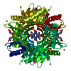
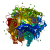
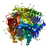
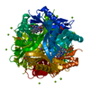
- Structure visualization
| Structure viewer | Molecule: MolmilJmol/JSmol |
|---|
- Downloads & links
-Download
| PDBx/mmCIF format | 4b2j.cif.gz | 31.4 KB | Display | PDBx/mmCIF format |
|---|---|---|---|---|
| PDB format | pdb4b2j.ent.gz | 21.4 KB | Display | PDB format |
| PDBx/mmJSON format | 4b2j.json.gz | Tree view | PDBx/mmJSON format | |
| Others |  Other downloads Other downloads |
-Validation report
| Arichive directory | https://data.pdbj.org/pub/pdb/validation_reports/b2/4b2jftp://data.pdbj.org/pub/pdb/validation_reports/b2/4b2j | HTTPS FTP |
|---|
-Related structure data
| Related structure data |  4b2kC  2cccS C: citing same article ( S: Starting model for refinement |
|---|---|
| Similar structure data |


-Links
 PDBj
PDBj- Assembly
| Deposited unit | 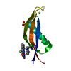
| ||||||||||||||||||||||||||||||||||||
|---|---|---|---|---|---|---|---|---|---|---|---|---|---|---|---|---|---|---|---|---|---|---|---|---|---|---|---|---|---|---|---|---|---|---|---|---|---|
| 1 | x 12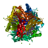
| ||||||||||||||||||||||||||||||||||||
| Unit cell |
| ||||||||||||||||||||||||||||||||||||
| Components on special symmetry positions |
|
-Components
-Protein , 1 types, 1 molecules A
| #1: Protein | Mass: 8531.203 Da / Num. of mol.: 1 Source method: isolated from a genetically manipulated source Details: TRYPTOPHAN 36 EXCHANGED BY 4F-FLUOROTRYPTOPHAN, RIBOFLAVIN BOUND Source: (gene. exp.) HALOBACTERIUM SALINARUM (Halophile) / Strain: R1 / Description: GERMAN COLLECTION OF MICROORGANISMS (DSM 671) / Plasmid: PQE80L / Production host:  ESCHERICHIA COLI (E. coli) / References: UniProt: B0R5M0 ESCHERICHIA COLI (E. coli) / References: UniProt: B0R5M0 |
|---|
-Non-polymers , 6 types, 93 molecules 


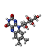

| #2: Chemical |  Mass: 24.305 Da / Num. of mol.: 2 / Source method: obtained synthetically / Formula: Mg Mass: 24.305 Da / Num. of mol.: 2 / Source method: obtained synthetically / Formula: Mg#3: Chemical | ChemComp-NA / |  Mass: 22.990 Da / Num. of mol.: 1 / Source method: obtained synthetically / Formula: Na Mass: 22.990 Da / Num. of mol.: 1 / Source method: obtained synthetically / Formula: Na#4: Chemical | ChemComp-CL / | Chloride Mass: 35.453 Da / Num. of mol.: 1 / Source method: obtained synthetically / Formula: Cl Mass: 35.453 Da / Num. of mol.: 1 / Source method: obtained synthetically / Formula: Cl#5: Chemical | ChemComp-SO4 / | Sulfate Mass: 96.063 Da / Num. of mol.: 1 / Source method: obtained synthetically / Formula: SO4 Mass: 96.063 Da / Num. of mol.: 1 / Source method: obtained synthetically / Formula: SO4#6: Chemical | ChemComp-RBF / | Riboflavin Mass: 376.364 Da / Num. of mol.: 1 / Source method: obtained synthetically / Formula: C17H20N4O6 Mass: 376.364 Da / Num. of mol.: 1 / Source method: obtained synthetically / Formula: C17H20N4O6#7: Water | ChemComp-HOH / | WaterMass: 18.015 Da / Num. of mol.: 87 / Source method: isolated from a natural source / Formula: H2O |
|---|
-Experimental details
-Experiment
| Experiment | Method: X-RAY DIFFRACTION / Number of used crystals: 2 |
|---|
- Sample preparation
| Crystal | Density Matthews: 3.64 Å3/Da / Density % sol: 66 % / Description: NONE |
|---|---|
| Crystal grow | pH: 7.5 Details: 0.1 M HEPES PH 7.5, PEG 400 30% (V/V), 2.5 M NACL, 0.2 M MGCL2 |
-Data collection
| Diffraction | Mean temperature: 100 K |
|---|---|
| Diffraction source | Source: SYNCHROTRON / Site: ESRF  / Beamline: ID23-2 / Wavelength: 0.873 / Beamline: ID23-2 / Wavelength: 0.873 |
| Detector | Type: MARMOSAIC 225 mm CCD / Detector: CCD / Date: Nov 20, 2009 |
| Radiation | Protocol: SINGLE WAVELENGTH / Monochromatic (M) / Laue (L): M / Scattering type: x-ray |
| Radiation wavelength | Wavelength: 0.873 Å / Relative weight: 1 |
| Reflection | Resolution: 1.9→20 Å / Num. obs: 10355 / % possible obs: 99.9 % / Observed criterion σ(I): 4 / Redundancy: 14 % / Rmerge(I) obs: 0.01 / Net I/σ(I): 20.8 |
| Reflection shell | Resolution: 1.9→2 Å / Redundancy: 14 % / Rmerge(I) obs: 0.6 / Mean I/σ(I) obs: 4.6 / % possible all: 100 |
- Processing
| Software |
| ||||||||||||||||||||||||||||||||||||||||||||||||||||||||||||||||||||||||||||||||||||||||||||||||||||||||||||||||||||||||||||||||||||||||||||||||||||||||||||||||||||||||||||||||||||||
|---|---|---|---|---|---|---|---|---|---|---|---|---|---|---|---|---|---|---|---|---|---|---|---|---|---|---|---|---|---|---|---|---|---|---|---|---|---|---|---|---|---|---|---|---|---|---|---|---|---|---|---|---|---|---|---|---|---|---|---|---|---|---|---|---|---|---|---|---|---|---|---|---|---|---|---|---|---|---|---|---|---|---|---|---|---|---|---|---|---|---|---|---|---|---|---|---|---|---|---|---|---|---|---|---|---|---|---|---|---|---|---|---|---|---|---|---|---|---|---|---|---|---|---|---|---|---|---|---|---|---|---|---|---|---|---|---|---|---|---|---|---|---|---|---|---|---|---|---|---|---|---|---|---|---|---|---|---|---|---|---|---|---|---|---|---|---|---|---|---|---|---|---|---|---|---|---|---|---|---|---|---|---|---|
| Refinement | Method to determine structure: MOLECULAR REPLACEMENT Starting model: PDB ENTRY 2CCC Resolution: 1.9→20 Å / Cor.coef. Fo:Fc: 0.956 / Cor.coef. Fo:Fc free: 0.958 / SU B: 1.82 / SU ML: 0.055 / Cross valid method: THROUGHOUT / ESU R: 0.093 / ESU R Free: 0.09 / Stereochemistry target values: MAXIMUM LIKELIHOOD Details: HYDROGENS HAVE BEEN ADDED IN THE RIDING POSITIONS. HYDROGENS HAVE BEEN USED IF PRESENT IN THE INPUT
| ||||||||||||||||||||||||||||||||||||||||||||||||||||||||||||||||||||||||||||||||||||||||||||||||||||||||||||||||||||||||||||||||||||||||||||||||||||||||||||||||||||||||||||||||||||||
| Solvent computation | Ion probe radii: 0.8 Å / Shrinkage radii: 0.8 Å / VDW probe radii: 1.2 Å / Solvent model: MASK | ||||||||||||||||||||||||||||||||||||||||||||||||||||||||||||||||||||||||||||||||||||||||||||||||||||||||||||||||||||||||||||||||||||||||||||||||||||||||||||||||||||||||||||||||||||||
| Displacement parameters | Biso mean: 24.198 Å2 | ||||||||||||||||||||||||||||||||||||||||||||||||||||||||||||||||||||||||||||||||||||||||||||||||||||||||||||||||||||||||||||||||||||||||||||||||||||||||||||||||||||||||||||||||||||||
| Refinement step | Cycle: LAST / Resolution: 1.9→20 Å
| ||||||||||||||||||||||||||||||||||||||||||||||||||||||||||||||||||||||||||||||||||||||||||||||||||||||||||||||||||||||||||||||||||||||||||||||||||||||||||||||||||||||||||||||||||||||
| Refine LS restraints |
|