 Movie
Movie Controller

[English] 日本語
 Yorodumi
- PDB-4anc: CRYSTAL FORM I OF THE D93N MUTANT OF NUCLEOSIDE DIPHOSPHATE KINAS... -
+ Open data

- Basic information
| Entry | Database: PDB / ID: 4anc | ||||||
|---|---|---|---|---|---|---|---|
| Title | CRYSTAL FORM I OF THE D93N MUTANT OF NUCLEOSIDE DIPHOSPHATE KINASE FROM MYCOBACTERIUM TUBERCULOSIS | ||||||
 Components Components | NUCLEOSIDE DIPHOSPHATE KINASE Nucleoside-diphosphate kinase Nucleoside-diphosphate kinase | ||||||
 Keywords Keywords | TRANSFERASE | ||||||
| Function / homology |  Function and homology information Function and homology informationsymbiont-mediated suppression of host innate immune response / : / purine nucleotide metabolic process / pyrimidine nucleotide metabolic process / nuclease activity / nucleoside-diphosphate kinase / UTP biosynthetic process / CTP biosynthetic process / GTP biosynthetic process / nucleoside diphosphate kinase activity ...symbiont-mediated suppression of host innate immune response / : / purine nucleotide metabolic process / pyrimidine nucleotide metabolic process / nuclease activity / nucleoside-diphosphate kinase / UTP biosynthetic process / CTP biosynthetic process / GTP biosynthetic process / nucleoside diphosphate kinase activity / phosphoprotein phosphatase activity / Prevention of phagosomal-lysosomal fusion / protein autophosphorylation / extracellular region / ATP binding / metal ion binding / plasma membrane / cytosol / cytoplasmSimilarity search - Function | ||||||
| Biological species |   MYCOBACTERIUM TUBERCULOSIS (bacteria) MYCOBACTERIUM TUBERCULOSIS (bacteria) | ||||||
| Method | X-RAY DIFFRACTION / SYNCHROTRON / MOLECULAR REPLACEMENT / Resolution: 2.8 Å | ||||||
 Authors Authors | Georgescauld, F. / Moynie, L. / Habersetzer, J. / Lascu, I. / Dautant, A. | ||||||
 Citation Citation | Journal: Plos One / Year: 2013 Title: Intersubunit Ionic Interactions Stabilize the Nucleoside Diphosphate Kinase of Mycobacterium Tuberculosis. Authors: Georgescauld, F. / Moynie, L. / Habersetzer, J. / Cervoni, L. / Mocan, I. / Borza, T. / Harris, P. / Dautant, A. / Lascu, I. #1: Journal: Proteins / Year: 2002Title: X-Ray Structure of Mycobacterium Tuberculosis Nucleoside Diphosphate Kinase. Authors: Chen, Y. / Morera, S. / Mocan, J. / Lascu, I. / Janin, J. | ||||||
| History |
|
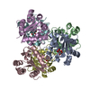
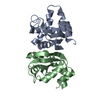
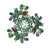
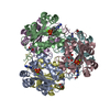
- Structure visualization
| Structure viewer | Molecule: MolmilJmol/JSmol |
|---|
- Downloads & links
-Download
| PDBx/mmCIF format | 4anc.cif.gz | 64.1 KB | Display | PDBx/mmCIF format |
|---|---|---|---|---|
| PDB format | pdb4anc.ent.gz | 48.3 KB | Display | PDB format |
| PDBx/mmJSON format | 4anc.json.gz | Tree view | PDBx/mmJSON format | |
| Others |  Other downloads Other downloads |
-Validation report
| Arichive directory | https://data.pdbj.org/pub/pdb/validation_reports/an/4ancftp://data.pdbj.org/pub/pdb/validation_reports/an/4anc | HTTPS FTP |
|---|
-Related structure data
| Related structure data |  4andC 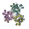 1k44S S: Starting model for refinement C: citing same article ( |
|---|---|
| Similar structure data |


-Links
 PDBj
PDBj

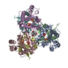
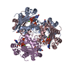
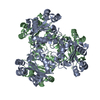
- Assembly
| Deposited unit | 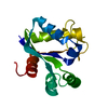
| ||||||||
|---|---|---|---|---|---|---|---|---|---|
| 1 | x 6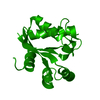
| ||||||||
| Unit cell |
| ||||||||
| Components on special symmetry positions |
|
-Components
| #1: Protein | Nucleoside-diphosphate kinase / NDK / NDKA / NDP KINASE / NUCLEOSIDE-2-P KINASE Mass: 14521.485 Da / Num. of mol.: 1 / Mutation: YES Source method: isolated from a genetically manipulated source Source: (gene. exp.) MYCOBACTERIUM TUBERCULOSIS (bacteria) / Production host: ESCHERICHIA COLI (E. coli) / Strain (production host): BL21(DE3) / Variant (production host): RILReferences: UniProt: P84284, UniProt: P9WJH7*PLUS, nucleoside-diphosphate kinase |
|---|---|
| #2: Water | ChemComp-HOH / Water Mass: 18.015 Da / Num. of mol.: 19 / Source method: isolated from a natural source / Formula: H2O Mass: 18.015 Da / Num. of mol.: 19 / Source method: isolated from a natural source / Formula: H2O |
| Compound details | ENGINEERED |
-Experimental details
-Experiment
| Experiment | Method: X-RAY DIFFRACTION / Number of used crystals: 1 |
|---|
- Sample preparation
| Crystal | Density Matthews: 3.93 Å3/Da / Density % sol: 68.5 % / Description: NONE |
|---|---|
| Crystal grow | pH: 8.5 / Details: 2.0 M AMMONIUM SULFATE, 0.1 M TRIS-HCL, PH 8.5 |
-Data collection
| Diffraction | Mean temperature: 107 K |
|---|---|
| Diffraction source | Source: SYNCHROTRON / Site: ESRF  / Beamline: ID23-2 / Wavelength: 0.873 / Beamline: ID23-2 / Wavelength: 0.873 |
| Detector | Type: MARMOSAIC 225 mm CCD / Detector: CCD / Date: Jul 15, 2006 |
| Radiation | Protocol: SINGLE WAVELENGTH / Monochromatic (M) / Laue (L): M / Scattering type: x-ray |
| Radiation wavelength | Wavelength: 0.873 Å / Relative weight: 1 |
| Reflection | Resolution: 2.8→26.9 Å / Num. obs: 6145 / % possible obs: 99.9 % / Observed criterion σ(I): 1 / Redundancy: 20.5 % / Biso Wilson estimate: 58.24 Å2 / Rmerge(I) obs: 0.09 / Net I/σ(I): 31.6 |
| Reflection shell | Resolution: 2.8→2.95 Å / Redundancy: 21 % / Rmerge(I) obs: 0.43 / Mean I/σ(I) obs: 7.3 / % possible all: 100 |
- Processing
| Software |
| ||||||||||||||||||||||||||||||||||||||||||||||||||||||||||||||||||||||||||||||||||||||||||||||||||||
|---|---|---|---|---|---|---|---|---|---|---|---|---|---|---|---|---|---|---|---|---|---|---|---|---|---|---|---|---|---|---|---|---|---|---|---|---|---|---|---|---|---|---|---|---|---|---|---|---|---|---|---|---|---|---|---|---|---|---|---|---|---|---|---|---|---|---|---|---|---|---|---|---|---|---|---|---|---|---|---|---|---|---|---|---|---|---|---|---|---|---|---|---|---|---|---|---|---|---|---|---|---|
| Refinement | Method to determine structure: MOLECULAR REPLACEMENT Starting model: PDB ENTRY 1K44 Resolution: 2.8→26.844 Å / SU ML: 0.36 / σ(F): 1.43 / Phase error: 24.58 / Stereochemistry target values: ML
| ||||||||||||||||||||||||||||||||||||||||||||||||||||||||||||||||||||||||||||||||||||||||||||||||||||
| Solvent computation | Shrinkage radii: 0.95 Å / VDW probe radii: 1.2 Å / Solvent model: FLAT BULK SOLVENT MODEL / Bsol: 43.685 Å2 / ksol: 0.323 e/Å3 | ||||||||||||||||||||||||||||||||||||||||||||||||||||||||||||||||||||||||||||||||||||||||||||||||||||
| Displacement parameters | Biso mean: 65.83 Å2
| ||||||||||||||||||||||||||||||||||||||||||||||||||||||||||||||||||||||||||||||||||||||||||||||||||||
| Refinement step | Cycle: LAST / Resolution: 2.8→26.844 Å
| ||||||||||||||||||||||||||||||||||||||||||||||||||||||||||||||||||||||||||||||||||||||||||||||||||||
| Refine LS restraints |
| ||||||||||||||||||||||||||||||||||||||||||||||||||||||||||||||||||||||||||||||||||||||||||||||||||||
| LS refinement shell |
| ||||||||||||||||||||||||||||||||||||||||||||||||||||||||||||||||||||||||||||||||||||||||||||||||||||
| Refinement TLS params. | Method: refined / Refine-ID: X-RAY DIFFRACTION
| ||||||||||||||||||||||||||||||||||||||||||||||||||||||||||||||||||||||||||||||||||||||||||||||||||||
| Refinement TLS group |
|
