 Movie
Movie Controller


+ Open data

- Basic information
| Entry | Database: PDB / ID: 4a3n | ||||||
|---|---|---|---|---|---|---|---|
| Title | Crystal Structure of HMG-BOX of Human SOX17 | ||||||
 Components Components | TRANSCRIPTION FACTOR SOX-17 | ||||||
 Keywords Keywords |  TRANSCRIPTION TRANSCRIPTION | ||||||
| Function / homology |  Function and homology information Function and homology informationcardiogenic plate morphogenesis / endodermal cell fate determination / regulation of cardiac cell fate specification / stem cell fate specification / endocardium formation / common bile duct development / endodermal digestive tract morphogenesis / ureter development / gallbladder development / inner cell mass cellular morphogenesis ...cardiogenic plate morphogenesis / endodermal cell fate determination / regulation of cardiac cell fate specification / stem cell fate specification / endocardium formation / common bile duct development / endodermal digestive tract morphogenesis / ureter development / gallbladder development / inner cell mass cellular morphogenesis / heart formation / cardiac cell fate determination / regulation of stem cell division / rostrocaudal neural tube patterning / endodermal cell fate specification / endoderm formation / cell migration involved in gastrulation / Specification of primordial germ cells / endocardial cell differentiation / positive regulation of endodermal cell differentiation / positive regulation of stem cell differentiation / embryonic foregut morphogenesis / regulation of stem cell proliferation / signal transduction involved in regulation of gene expression / Formation of definitive endoderm / metanephros development / embryonic heart tube development / embryonic heart tube morphogenesis / heart looping / outflow tract morphogenesis / regulation of embryonic development / anatomical structure morphogenesis / vasculogenesis / cellular response to leukemia inhibitory factor / Deactivation of the beta-catenin transactivating complex / negative regulation of canonical Wnt signaling pathway / protein destabilization / negative regulation of cell growth / beta-catenin binding / Wnt signaling pathway / positive regulation of protein catabolic process / gene expression / heart development / spermatogenesis / DNA-binding transcription activator activity, RNA polymerase II-specific / angiogenesis / transcription regulator complex / RNA polymerase II-specific DNA-binding transcription factor binding / protein stabilization / transcription cis-regulatory region binding / DNA-binding transcription factor activity, RNA polymerase II-specific / RNA polymerase II cis-regulatory region sequence-specific DNA binding / chromatin / positive regulation of gene expression / regulation of DNA-templated transcription / regulation of transcription by RNA polymerase II / positive regulation of DNA-templated transcription / positive regulation of transcription by RNA polymerase II / nucleoplasm / nucleusSimilarity search - Function | ||||||
| Biological species |  HOMO SAPIENS (human) HOMO SAPIENS (human) | ||||||
| Method | X-RAY DIFFRACTION / SYNCHROTRON / MOLECULAR REPLACEMENT / Resolution: 2.4 Å | ||||||
 Authors Authors | Gao, N. / Gao, H. / Qian, H. / Si, S. / Xie, Y. | ||||||
 Citation Citation | Journal: Protein Pept.Lett. / Year: 2013 Title: Structural Basis of Human Transcription Factor Sry-Related Box 17 Binding to DNA. Authors: Gao, N. / Jiang, W. / Gao, H. / Cheng, Z. / Qian, H. / Si, S. / Xie, Y. #1: Journal: J.Mol.Biol. / Year: 2009Title: The Structure of Sox17 Bound to DNA Reveals a Conserved Bending Topology But Selective Protein Interaction Platforms. Authors: Palasingam, P. / Jauch, R. / Ng, C.K.L. / Kolatkar, P.R. | ||||||
| History |
|
- Structure visualization
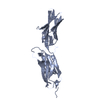
| Structure viewer | Molecule: MolmilJmol/JSmol |
|---|
- Downloads & links
-Download
| PDBx/mmCIF format | 4a3n.cif.gz | 28.9 KB | Display | PDBx/mmCIF format |
|---|---|---|---|---|
| PDB format | pdb4a3n.ent.gz | 17.9 KB | Display | PDB format |
| PDBx/mmJSON format | 4a3n.json.gz | Tree view | PDBx/mmJSON format | |
| Others |  Other downloads Other downloads |
-Validation report
| Arichive directory | https://data.pdbj.org/pub/pdb/validation_reports/a3/4a3nftp://data.pdbj.org/pub/pdb/validation_reports/a3/4a3n | HTTPS FTP |
|---|
-Related structure data
| Related structure data | 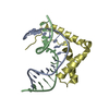 3f27S S: Starting model for refinement |
|---|---|
| Similar structure data |


-Links
 PDBj
PDBj

- Assembly
| Deposited unit | 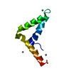
| ||||||||
|---|---|---|---|---|---|---|---|---|---|
| 1 | 
| ||||||||
| Unit cell |
|
-Components
| #1: Protein | Mass: 8445.764 Da / Num. of mol.: 1 / Fragment: HMG-BOX, RESIDUES 68-136 Source method: isolated from a genetically manipulated source Source: (gene. exp.) HOMO SAPIENS (human) / Production host:  ESCHERICHIA COLI (E. coli) / Strain (production host): BL21(DE3) / References: UniProt: Q9H6I2 ESCHERICHIA COLI (E. coli) / Strain (production host): BL21(DE3) / References: UniProt: Q9H6I2 | ||
|---|---|---|---|
| #2: Chemical |   Mass: 65.409 Da / Num. of mol.: 3 / Source method: obtained synthetically / Formula: Zn Mass: 65.409 Da / Num. of mol.: 3 / Source method: obtained synthetically / Formula: Zn#3: Water | ChemComp-HOH / | Water Mass: 18.015 Da / Num. of mol.: 72 / Source method: isolated from a natural source / Formula: H2O Mass: 18.015 Da / Num. of mol.: 72 / Source method: isolated from a natural source / Formula: H2O |
-Experimental details
-Experiment
| Experiment | Method: X-RAY DIFFRACTION |
|---|
- Sample preparation
| Crystal | Density Matthews: 3.25 Å3/Da / Density % sol: 62.19 % / Description: NONE |
|---|
-Data collection
| Diffraction | Mean temperature: 100 K |
|---|---|
| Diffraction source | Source: SYNCHROTRON / Site: Photon Factory  / Beamline: BL-5A / Wavelength: 0.979 / Beamline: BL-5A / Wavelength: 0.979 |
| Detector | Type: ADSC QUANTUM 315r / Detector: CCD |
| Radiation | Protocol: SINGLE WAVELENGTH / Monochromatic (M) / Laue (L): M / Scattering type: x-ray |
| Radiation wavelength | Wavelength: 0.979 Å / Relative weight: 1 |
| Reflection | Resolution: 2.4→50 Å / Num. obs: 4389 / % possible obs: 98 % / Observed criterion σ(I): 0 / Redundancy: 3.4 % / Biso Wilson estimate: 20.9 Å2 / Rmerge(I) obs: 0.08 / Net I/σ(I): 22.9 |
| Reflection shell | Resolution: 2.4→2.53 Å / Redundancy: 3.5 % / Rmerge(I) obs: 0.19 / Mean I/σ(I) obs: 23.4 / % possible all: 100 |
- Processing
| Software |
| ||||||||||||||||||||||||||||||||||||||||||||||||||||||||||||
|---|---|---|---|---|---|---|---|---|---|---|---|---|---|---|---|---|---|---|---|---|---|---|---|---|---|---|---|---|---|---|---|---|---|---|---|---|---|---|---|---|---|---|---|---|---|---|---|---|---|---|---|---|---|---|---|---|---|---|---|---|---|
| Refinement | Method to determine structure: MOLECULAR REPLACEMENT Starting model: PDB ENTRY 3F27 Resolution: 2.4→23.66 Å / Rfactor Rfree error: 0.013 / Data cutoff high absF: 1313405.55 / Data cutoff low absF: 0 / Isotropic thermal model: RESTRAINED / Cross valid method: THROUGHOUT / σ(F): 2 / Details: BULK SOLVENT MODEL USED
| ||||||||||||||||||||||||||||||||||||||||||||||||||||||||||||
| Solvent computation | Solvent model: FLAT MODEL / Bsol: 60.9136 Å2 / ksol: 0.35 e/Å3 | ||||||||||||||||||||||||||||||||||||||||||||||||||||||||||||
| Displacement parameters | Biso mean: 44.7 Å2
| ||||||||||||||||||||||||||||||||||||||||||||||||||||||||||||
| Refine analyze |
| ||||||||||||||||||||||||||||||||||||||||||||||||||||||||||||
| Refinement step | Cycle: LAST / Resolution: 2.4→23.66 Å
| ||||||||||||||||||||||||||||||||||||||||||||||||||||||||||||
| Refine LS restraints |
| ||||||||||||||||||||||||||||||||||||||||||||||||||||||||||||
| Refine LS restraints NCS | NCS model details: NONE | ||||||||||||||||||||||||||||||||||||||||||||||||||||||||||||
| LS refinement shell | Resolution: 2.4→2.55 Å / Rfactor Rfree error: 0.046 / Total num. of bins used: 6
| ||||||||||||||||||||||||||||||||||||||||||||||||||||||||||||
| Xplor file |
|
