 Movie
Movie Controller

[English] 日本語
 Yorodumi
- PDB-3ztp: Orthorhombic crystal form P21212 of the Aquifex aeolicus nucleosi... -
+ Open data

- Basic information
| Entry | Database: PDB / ID: 3ztp | |||||||||
|---|---|---|---|---|---|---|---|---|---|---|
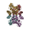
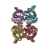
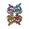
| Title | Orthorhombic crystal form P21212 of the Aquifex aeolicus nucleoside diphosphate kinase | |||||||||
 Components Components | NUCLEOSIDE DIPHOSPHATE KINASE Nucleoside-diphosphate kinase Nucleoside-diphosphate kinase | |||||||||
 Keywords Keywords | TRANSFERASE | |||||||||
| Function / homology |  Function and homology information Function and homology informationpurine nucleotide metabolic process / pyrimidine nucleotide metabolic process / nucleoside-diphosphate kinase / UTP biosynthetic process / CTP biosynthetic process / GTP biosynthetic process / nucleoside diphosphate kinase activity / phosphorylation / ATP binding / metal ion binding / cytoplasmSimilarity search - Function | |||||||||
| Biological species |   AQUIFEX AEOLICUS (bacteria) AQUIFEX AEOLICUS (bacteria) | |||||||||
| Method | X-RAY DIFFRACTION / SYNCHROTRON / MOLECULAR REPLACEMENT / Resolution: 1.37 Å | |||||||||
 Authors Authors | Boissier, F. / Georgescauld, F. / Moynie, L. / Dupuy, J.-W. / Sarger, C. / Podar, M. / Lascu, I. / Giraud, M.-F. / Dautant, A. | |||||||||
 Citation Citation | Journal: Proteins / Year: 2012 Title: An Intersubunit Disulfide Bridge Stabilizes the Tetrameric Nucleoside Diphosphate Kinase of Aquifex Aeolicus. Authors: Boissier, F. / Georgescauld, F. / Moynie, L. / Dupuy, J.W. / Sarger, C. / Podar, M. / Lascu, I. / Giraud, M.F. / Dautant, A. #1: Journal: Proteins / Year: 2007Title: The Structure of the Escherichia Coli Nucleoside Diphosphate Kinase Reveals a New Quaternary Architecture for This Enzyme Family. Authors: Moynie, L. / Giraud, M. / Georgescauld, F. / Lascu, I. / Dautant, A. #2: Journal: J.Mol.Biol. / Year: 1993Title: Crystal Structure of Myxococcus Xanthus Nucleoside Diphosphate Kinase and its Interaction with a Nucleotide Substrate at 2.0 A Resolution. Authors: Williams, R.L. / Oren, D.A. / Munoz-Dorado, J. / Inouye, S. / Inouye, M. / Arnold, E. | |||||||||
| History |
|
- Structure visualization
| Structure viewer | Molecule: MolmilJmol/JSmol |
|---|
- Downloads & links
-Download
| PDBx/mmCIF format | 3ztp.cif.gz | 188.4 KB | Display | PDBx/mmCIF format |
|---|---|---|---|---|
| PDB format | pdb3ztp.ent.gz | 154.2 KB | Display | PDB format |
| PDBx/mmJSON format | 3ztp.json.gz | Tree view | PDBx/mmJSON format | |
| Others |  Other downloads Other downloads |
-Validation report
| Arichive directory | https://data.pdbj.org/pub/pdb/validation_reports/zt/3ztpftp://data.pdbj.org/pub/pdb/validation_reports/zt/3ztp | HTTPS FTP |
|---|
-Related structure data
| Related structure data | 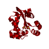 3ztoSC 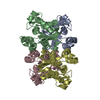 3ztqC  3ztrC  3ztsC C: citing same article ( S: Starting model for refinement |
|---|---|
| Similar structure data |

-Links
 PDBj
PDBj- Assembly
| Deposited unit | 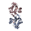
| |||||||||
|---|---|---|---|---|---|---|---|---|---|---|
| 1 | 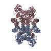
| |||||||||
| Unit cell |
| |||||||||
| Components on special symmetry positions |
|
-Components
| #1: Protein | Nucleoside-diphosphate kinase / NDK / NDP KINASE / NUCLEOSIDE-2-P KINASE Mass: 15963.325 Da / Num. of mol.: 2 Source method: isolated from a genetically manipulated source Source: (gene. exp.) AQUIFEX AEOLICUS (bacteria) / Production host: ESCHERICHIA COLI (E. coli) / Strain (production host): BL21(DE3) / References: UniProt: O67528, nucleoside-diphosphate kinase#2: Chemical | ChemComp-GOL / | Glycerol  Mass: 92.094 Da / Num. of mol.: 1 / Source method: obtained synthetically / Formula: C3H8O3 Mass: 92.094 Da / Num. of mol.: 1 / Source method: obtained synthetically / Formula: C3H8O3#3: Chemical | Sulfate  Mass: 96.063 Da / Num. of mol.: 2 / Source method: obtained synthetically / Formula: SO4 Mass: 96.063 Da / Num. of mol.: 2 / Source method: obtained synthetically / Formula: SO4#4: Water | ChemComp-HOH / | Water Mass: 18.015 Da / Num. of mol.: 356 / Source method: isolated from a natural source / Formula: H2O Mass: 18.015 Da / Num. of mol.: 356 / Source method: isolated from a natural source / Formula: H2O |
|---|
-Experimental details
-Experiment
| Experiment | Method: X-RAY DIFFRACTION / Number of used crystals: 1 |
|---|
- Sample preparation
| Crystal | Density Matthews: 2.44 Å3/Da / Density % sol: 49.7 % / Description: NONE |
|---|---|
| Crystal grow | pH: 7.5 / Details: 2.0 M AMMONIUM FORMATE, 0.1 M HEPES, PH 7.5 |
-Data collection
| Diffraction | Mean temperature: 100 K |
|---|---|
| Diffraction source | Source: SYNCHROTRON / Site: ESRF  / Beamline: ID14-1 / Wavelength: 0.9334 / Beamline: ID14-1 / Wavelength: 0.9334 |
| Detector | Type: ADSC QUANTUM 315r / Detector: CCD / Date: Feb 6, 2009 / Details: MIRRORS |
| Radiation | Monochromator: DIAMOND (111), GE(220) / Protocol: SINGLE WAVELENGTH / Monochromatic (M) / Laue (L): M / Scattering type: x-ray |
| Radiation wavelength | Wavelength: 0.9334 Å / Relative weight: 1 |
| Reflection | Resolution: 1.37→23.31 Å / Num. obs: 65505 / % possible obs: 97.8 % / Observed criterion σ(I): 1 / Redundancy: 3.3 % / Biso Wilson estimate: 11.45 Å2 / Rmerge(I) obs: 0.06 / Net I/σ(I): 12.4 |
| Reflection shell | Resolution: 1.37→1.43 Å / Redundancy: 2.1 % / Rmerge(I) obs: 0.31 / Mean I/σ(I) obs: 2.5 / % possible all: 97.8 |
- Processing
| Software |
| |||||||||||||||||||||||||||||||||||||||||||||||||||||||||||||||||||||||||||||||||||||||||||||||||||||||||||||||||||||||||||||
|---|---|---|---|---|---|---|---|---|---|---|---|---|---|---|---|---|---|---|---|---|---|---|---|---|---|---|---|---|---|---|---|---|---|---|---|---|---|---|---|---|---|---|---|---|---|---|---|---|---|---|---|---|---|---|---|---|---|---|---|---|---|---|---|---|---|---|---|---|---|---|---|---|---|---|---|---|---|---|---|---|---|---|---|---|---|---|---|---|---|---|---|---|---|---|---|---|---|---|---|---|---|---|---|---|---|---|---|---|---|---|---|---|---|---|---|---|---|---|---|---|---|---|---|---|---|---|
| Refinement | Method to determine structure: MOLECULAR REPLACEMENT Starting model: PDB ENTRY 3ZTO Resolution: 1.37→23.313 Å / SU ML: 0.14 / σ(F): 0 / Phase error: 13.7 / Stereochemistry target values: ML Details: IN THE SSBOND CARDS: THE FIRST ATOM BELONGS TO THE ALTERNATE CONFORMER A (WITH OCCUPANCY Q1). THE SECOND ONE BELONGS TO THE ALTERNATE CONFORMER B (WITH OCCUPANCY Q2). ABS(Q1 - Q2)*100 IS THE ...Details: IN THE SSBOND CARDS: THE FIRST ATOM BELONGS TO THE ALTERNATE CONFORMER A (WITH OCCUPANCY Q1). THE SECOND ONE BELONGS TO THE ALTERNATE CONFORMER B (WITH OCCUPANCY Q2). ABS(Q1 - Q2)*100 IS THE PERCENTAGE OF CYS133 NOT INVOLVED IN THE DISULFIDE BRIDGE.
| |||||||||||||||||||||||||||||||||||||||||||||||||||||||||||||||||||||||||||||||||||||||||||||||||||||||||||||||||||||||||||||
| Solvent computation | Shrinkage radii: 0.41 Å / VDW probe radii: 0.6 Å / Solvent model: FLAT BULK SOLVENT MODEL / Bsol: 61.221 Å2 / ksol: 0.478 e/Å3 | |||||||||||||||||||||||||||||||||||||||||||||||||||||||||||||||||||||||||||||||||||||||||||||||||||||||||||||||||||||||||||||
| Displacement parameters | Biso mean: 17.5 Å2
| |||||||||||||||||||||||||||||||||||||||||||||||||||||||||||||||||||||||||||||||||||||||||||||||||||||||||||||||||||||||||||||
| Refinement step | Cycle: LAST / Resolution: 1.37→23.313 Å
| |||||||||||||||||||||||||||||||||||||||||||||||||||||||||||||||||||||||||||||||||||||||||||||||||||||||||||||||||||||||||||||
| Refine LS restraints |
| |||||||||||||||||||||||||||||||||||||||||||||||||||||||||||||||||||||||||||||||||||||||||||||||||||||||||||||||||||||||||||||
| LS refinement shell |
| |||||||||||||||||||||||||||||||||||||||||||||||||||||||||||||||||||||||||||||||||||||||||||||||||||||||||||||||||||||||||||||
| Refinement TLS params. | Method: refined / Refine-ID: X-RAY DIFFRACTION
| |||||||||||||||||||||||||||||||||||||||||||||||||||||||||||||||||||||||||||||||||||||||||||||||||||||||||||||||||||||||||||||
| Refinement TLS group |
|
