 Movie
Movie Controller

[English] 日本語
 Yorodumi
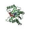
- PDB-3zfq: Crystal structure of product-like, processed N-terminal protease ... -
+ Open data

- Basic information
| Entry | Database: PDB / ID: 3zfq | ||||||
|---|---|---|---|---|---|---|---|
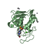
| Title | Crystal structure of product-like, processed N-terminal protease Npro with mercury | ||||||
 Components Components | N-TERMINAL PROTEASE NPRO | ||||||
 Keywords Keywords |  HYDROLASE / AUTO-PROCESSING CYSTEINE PROTEASE / VIRAL PROTEASE / IN CIS- CLEAVAGE / HYDROXIDE-DEPENDENT CATALYSIS / AUTO-PROTEOLYSIS / IMMUNE MODULATION / HOST-PATHOGEN INTERACTION / CONVERGENT EVOLUTION HYDROLASE / AUTO-PROCESSING CYSTEINE PROTEASE / VIRAL PROTEASE / IN CIS- CLEAVAGE / HYDROXIDE-DEPENDENT CATALYSIS / AUTO-PROTEOLYSIS / IMMUNE MODULATION / HOST-PATHOGEN INTERACTION / CONVERGENT EVOLUTION | ||||||
| Function / homology |  Function and homology information Function and homology informationsymbiont-mediated suppression of host cytoplasmic pattern recognition receptor signaling pathway via inhibition of IRF3 activity / viral protein processing / cysteine-type endopeptidase activity / proteolysisSimilarity search - Function | ||||||
| Biological species |  PESTIVIRUS STRAIN D32/00_HOBI PESTIVIRUS STRAIN D32/00_HOBI | ||||||
| Method | X-RAY DIFFRACTION / MOLECULAR REPLACEMENT / Resolution: 2.65 Å | ||||||
 Authors Authors | Zogg, T. / Sponring, M. / Schindler, S. / Koll, M. / Schneider, R. / Brandstetter, H. / Auer, B. | ||||||
 Citation Citation | Journal: Structure / Year: 2013 Title: Crystal Structures of the Viral Protease Npro Imply Distinct Roles for the Catalytic Water in Catalysis Authors: Zogg, T. / Sponring, M. / Schindler, S. / Koll, M. / Schneider, R. / Brandstetter, H. / Auer, B. | ||||||
| History |
|
- Structure visualization
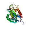
| Structure viewer | Molecule: MolmilJmol/JSmol |
|---|
- Downloads & links
-Download
| PDBx/mmCIF format | 3zfq.cif.gz | 43.9 KB | Display | PDBx/mmCIF format |
|---|---|---|---|---|
| PDB format | pdb3zfq.ent.gz | 29.4 KB | Display | PDB format |
| PDBx/mmJSON format | 3zfq.json.gz | Tree view | PDBx/mmJSON format | |
| Others |  Other downloads Other downloads |
-Validation report
| Arichive directory | https://data.pdbj.org/pub/pdb/validation_reports/zf/3zfqftp://data.pdbj.org/pub/pdb/validation_reports/zf/3zfq | HTTPS FTP |
|---|
-Related structure data
| Related structure data | 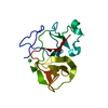 3zfnSC 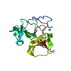 3zfoC 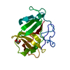 3zfpC 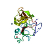 3zfrC  3zftC 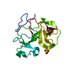 3zfuC S: Starting model for refinement C: citing same article ( |
|---|---|
| Similar structure data |


-Links
 PDBj
PDBj


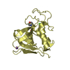
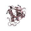
- Assembly
| Deposited unit | 
| ||||||||
|---|---|---|---|---|---|---|---|---|---|
| 1 |
| ||||||||
| Unit cell |
|
-Components
| #1: Protein | Mass: 16558.031 Da / Num. of mol.: 1 / Fragment: NPRO, RESIDUES 22-168 / Mutation: YES Source method: isolated from a genetically manipulated source Source: (gene. exp.) PESTIVIRUS STRAIN D32/00_HOBI / Production host:  ESCHERICHIA COLI (E. coli) / Strain (production host): BL21(DE3) ESCHERICHIA COLI (E. coli) / Strain (production host): BL21(DE3)References: UniProt: Q5L4B1, Hydrolases; Acting on peptide bonds (peptidases); Cysteine endopeptidases |
|---|---|
| #2: Chemical | ChemComp-SGM / 3-Mercaptopropane-1,2-diol  Mass: 108.159 Da / Num. of mol.: 1 / Source method: obtained synthetically / Formula: C3H8O2S Mass: 108.159 Da / Num. of mol.: 1 / Source method: obtained synthetically / Formula: C3H8O2S |
| #3: Chemical | ChemComp-HG / Mercury (element)  Mass: 200.590 Da / Num. of mol.: 1 / Source method: obtained synthetically / Formula: Hg Mass: 200.590 Da / Num. of mol.: 1 / Source method: obtained synthetically / Formula: Hg |
| #4: Water | ChemComp-HOH / Water Mass: 18.015 Da / Num. of mol.: 23 / Source method: isolated from a natural source / Formula: H2O Mass: 18.015 Da / Num. of mol.: 23 / Source method: isolated from a natural source / Formula: H2O |
-Experimental details
-Experiment
| Experiment | Method: X-RAY DIFFRACTION / Number of used crystals: 1 |
|---|
- Sample preparation
| Crystal | Density Matthews: 2.1 Å3/Da / Density % sol: 42 % / Description: NONE |
|---|---|
| Crystal grow | pH: 8.5 / Details: 100MM NAACETATE, PH 8.5 50% PEG6000 |
-Data collection
| Diffraction | Mean temperature: 100 K |
|---|---|
| Diffraction source | Source: ROTATING ANODE / Type: BRUKER AXS MICROSTAR / Wavelength: 1.5418 |
| Detector | Type: MARRESEARCH / Detector: IMAGE PLATE / Date: Mar 14, 2011 |
| Radiation | Protocol: SINGLE WAVELENGTH / Monochromatic (M) / Laue (L): M / Scattering type: x-ray |
| Radiation wavelength | Wavelength: 1.5418 Å / Relative weight: 1 |
| Reflection | Resolution: 2.6→10 Å / Num. obs: 4228 / % possible obs: 100 % / Observed criterion σ(I): -3 / Redundancy: 3.5 % / Rmerge(I) obs: 0.14 / Net I/σ(I): 7.6 |
| Reflection shell | Resolution: 2.6→2.74 Å / Redundancy: 3.5 % / Rmerge(I) obs: 0.45 / Mean I/σ(I) obs: 2.8 / % possible all: 100 |
- Processing
| Software |
| ||||||||||||||||||||||||||||||||||||||||||||||||||||||||||||||||||||||||||||||||||||||||||||||||||||||||||||||||||||||||||||||||||||||||||||||||||||||||||||||||||||||||||||||||||||||
|---|---|---|---|---|---|---|---|---|---|---|---|---|---|---|---|---|---|---|---|---|---|---|---|---|---|---|---|---|---|---|---|---|---|---|---|---|---|---|---|---|---|---|---|---|---|---|---|---|---|---|---|---|---|---|---|---|---|---|---|---|---|---|---|---|---|---|---|---|---|---|---|---|---|---|---|---|---|---|---|---|---|---|---|---|---|---|---|---|---|---|---|---|---|---|---|---|---|---|---|---|---|---|---|---|---|---|---|---|---|---|---|---|---|---|---|---|---|---|---|---|---|---|---|---|---|---|---|---|---|---|---|---|---|---|---|---|---|---|---|---|---|---|---|---|---|---|---|---|---|---|---|---|---|---|---|---|---|---|---|---|---|---|---|---|---|---|---|---|---|---|---|---|---|---|---|---|---|---|---|---|---|---|---|
| Refinement | Method to determine structure: MOLECULAR REPLACEMENT Starting model: PDB ENTRY 3ZFN Resolution: 2.65→10 Å / Cor.coef. Fo:Fc: 0.91 / Cor.coef. Fo:Fc free: 0.833 / SU B: 11.742 / SU ML: 0.262 / Cross valid method: THROUGHOUT / ESU R Free: 0.415 / Stereochemistry target values: MAXIMUM LIKELIHOOD Details: HYDROGENS HAVE BEEN ADDED IN THE RIDING POSITIONS. RESIDUES 145-150 DISORDERED
| ||||||||||||||||||||||||||||||||||||||||||||||||||||||||||||||||||||||||||||||||||||||||||||||||||||||||||||||||||||||||||||||||||||||||||||||||||||||||||||||||||||||||||||||||||||||
| Solvent computation | Ion probe radii: 0.8 Å / Shrinkage radii: 0.8 Å / VDW probe radii: 1.2 Å / Solvent model: MASK | ||||||||||||||||||||||||||||||||||||||||||||||||||||||||||||||||||||||||||||||||||||||||||||||||||||||||||||||||||||||||||||||||||||||||||||||||||||||||||||||||||||||||||||||||||||||
| Displacement parameters | Biso mean: 13.94 Å2
| ||||||||||||||||||||||||||||||||||||||||||||||||||||||||||||||||||||||||||||||||||||||||||||||||||||||||||||||||||||||||||||||||||||||||||||||||||||||||||||||||||||||||||||||||||||||
| Refinement step | Cycle: LAST / Resolution: 2.65→10 Å
| ||||||||||||||||||||||||||||||||||||||||||||||||||||||||||||||||||||||||||||||||||||||||||||||||||||||||||||||||||||||||||||||||||||||||||||||||||||||||||||||||||||||||||||||||||||||
| Refine LS restraints |
|