 Movie
Movie Controller

[English] 日本語
 Yorodumi
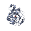
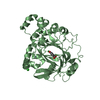
- PDB-3ze4: Crystal structure of the integral membrane diacylglycerol kinase ... -
+ Open data

- Basic information
| Entry | Database: PDB / ID: 3ze4 | ||||||
|---|---|---|---|---|---|---|---|
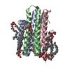
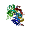
| Title | Crystal structure of the integral membrane diacylglycerol kinase - wild-type | ||||||
 Components Components | DIACYLGLYCEROL KINASE | ||||||
 Keywords Keywords | TRANSFERASE / LIPID METABOLISM / IN MESO CRYSTALLISATION / LIPID CUBIC PHASE / LIPIDIC MESOPHASE / MEMBRANE PROTEIN / MONOACYLGLYCEROL / 7.8 MAG | ||||||
| Function / homology |  Function and homology information Function and homology informationdiacylglycerol kinase (ATP) / ATP-dependent diacylglycerol kinase activity / phosphatidic acid biosynthetic process / response to UV / phosphorylation / ATP binding / membrane / identical protein binding / metal ion binding / plasma membraneSimilarity search - Function | ||||||
| Biological species |  ESCHERICHIA COLI K-12 (bacteria) ESCHERICHIA COLI K-12 (bacteria) | ||||||
| Method | X-RAY DIFFRACTION / SYNCHROTRON / MOLECULAR REPLACEMENT / Resolution: 3.702 Å | ||||||
| Model type details | P ATOMS ONLY, CHAIN D, E, F | ||||||
 Authors Authors | Li, D. / Lyons, J.A. / Pye, V.E. / Vogeley, L. / Aragao, D. / Caffrey, M. | ||||||
 Citation Citation | Journal: Nature / Year: 2013 Title: Crystal Structure of the Integral Membrane Diacylglycerol Kinase. Authors: Li, D. / Lyons, J.A. / Pye, V.E. / Vogeley, L. / Aragao, D. / Kenyon, C.P. / Shah, S.T.A. / Doherty, C. / Aherne, M. / Caffrey, M. | ||||||
| History |
|
- Structure visualization
| Structure viewer | Molecule: MolmilJmol/JSmol |
|---|
- Downloads & links
-Download
| PDBx/mmCIF format | 3ze4.cif.gz | 136.9 KB | Display | PDBx/mmCIF format |
|---|---|---|---|---|
| PDB format | pdb3ze4.ent.gz | 111.9 KB | Display | PDB format |
| PDBx/mmJSON format | 3ze4.json.gz | Tree view | PDBx/mmJSON format | |
| Others |  Other downloads Other downloads |
-Validation report
| Arichive directory | https://data.pdbj.org/pub/pdb/validation_reports/ze/3ze4ftp://data.pdbj.org/pub/pdb/validation_reports/ze/3ze4 | HTTPS FTP |
|---|
-Related structure data
| Related structure data | 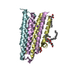 3ze3C  3ze5SC C: citing same article ( S: Starting model for refinement |
|---|---|
| Similar structure data |


-Links
 PDBj
PDBj- Assembly
| Deposited unit | 
| ||||||||||||||||||||||||||||||||||||
|---|---|---|---|---|---|---|---|---|---|---|---|---|---|---|---|---|---|---|---|---|---|---|---|---|---|---|---|---|---|---|---|---|---|---|---|---|---|
| 1 |
| ||||||||||||||||||||||||||||||||||||
| Unit cell |
| ||||||||||||||||||||||||||||||||||||
| Noncrystallographic symmetry (NCS) | NCS domain:
NCS domain segments:
NCS oper:
|
-Components
| #1: Protein | / DAGK / DIGLYCERIDE KINASE / DGK Mass: 14252.625 Da / Num. of mol.: 3 Source method: isolated from a genetically manipulated source Source: (gene. exp.) ESCHERICHIA COLI K-12 (bacteria)Description: THE WILD-TYPE GENE WAS SYNTHESIZED BASED ON THE DGKA NUCLEOTIDE SEQUENCE OF ESCHERICHIA COLI K12, WITH ADDITIONAL NUCLEOTIDES ENCODING HIS TAG SEQUENCES AT THE N-TERMINUS. Plasmid: PTRCHISB_DGKA_WILD-TYPE / Production host: ESCHERICHIA COLI (E. coli) / Strain (production host): WH1061 / References: UniProt: P0ABN1, diacylglycerol kinase (ATP) |
|---|
-Experimental details
-Experiment
| Experiment | Method: X-RAY DIFFRACTION / Number of used crystals: 1 |
|---|
- Sample preparation
| Crystal | Density Matthews: 3.74 Å3/Da / Density % sol: 67.16 % / Description: NONE |
|---|---|
| Crystal grow | Temperature: 277 K / Method: lipidic cubic phase / pH: 5.6 Details: 3-5 %(V/V) 2-METHYL-2, 4-PENTANEDIOL, 0.1 M SODIUM CHLORIDE, 0.1 M LITHIUM NITRATE, 0.1 M SODIUM CITRATE/HCL PH 5.6. CRYSTALLIZED USING THE IN MESO (LIPIDIC CUBIC PHASE) METHOD AT 4 DEGREE ...Details: 3-5 %(V/V) 2-METHYL-2, 4-PENTANEDIOL, 0.1 M SODIUM CHLORIDE, 0.1 M LITHIUM NITRATE, 0.1 M SODIUM CITRATE/HCL PH 5.6. CRYSTALLIZED USING THE IN MESO (LIPIDIC CUBIC PHASE) METHOD AT 4 DEGREE CELSIUS WITH THE 7.8 MONOACYLGLYCEROL (7.8 MAG) AS THE HOSTING LIPID. |
-Data collection
| Diffraction | Mean temperature: 100 K |
|---|---|
| Diffraction source | Source: SYNCHROTRON / Site: APS  / Beamline: 23-ID-B / Wavelength: 1.03306 / Beamline: 23-ID-B / Wavelength: 1.03306 |
| Detector | Type: MARRESEARCH / Detector: CCD / Date: Jun 13, 2012 / Details: K-B PAIR OF BIOMORPH MIRRORS |
| Radiation | Monochromator: SI(111) DOUBLE CRYSTAL / Protocol: SINGLE WAVELENGTH / Monochromatic (M) / Laue (L): M / Scattering type: x-ray |
| Radiation wavelength | Wavelength: 1.03306 Å / Relative weight: 1 |
| Reflection | Resolution: 3.7→39.23 Å / Num. obs: 7069 / % possible obs: 96.7 % / Observed criterion σ(I): -3 / Redundancy: 4.8 % / Biso Wilson estimate: 130.23 Å2 / Rmerge(I) obs: 0.08 / Net I/σ(I): 11.8 |
| Reflection shell | Resolution: 3.7→3.8 Å / Redundancy: 4.7 % / Rmerge(I) obs: 0.72 / Mean I/σ(I) obs: 2 / % possible all: 98 |
- Processing
| Software |
| ||||||||||||||||||||||||||||||||||||||||||||||||||||||||||||||||||||||||||||||||||||||||||||||||||||||||||||||||||||||||||||||||||||||||||||||||||||||||||||||||||||||||||||||||||||||||||||||||||||||||||||||||||||||||||||||||||||||||||||||||||||||||||||||||||||||||||||||||||||||||||||||||||||||||||||||||||||||||||||||||||||||||||||||||||||||||||||||||||||||||||||||||||||||||||||||||||||||||||||||||
|---|---|---|---|---|---|---|---|---|---|---|---|---|---|---|---|---|---|---|---|---|---|---|---|---|---|---|---|---|---|---|---|---|---|---|---|---|---|---|---|---|---|---|---|---|---|---|---|---|---|---|---|---|---|---|---|---|---|---|---|---|---|---|---|---|---|---|---|---|---|---|---|---|---|---|---|---|---|---|---|---|---|---|---|---|---|---|---|---|---|---|---|---|---|---|---|---|---|---|---|---|---|---|---|---|---|---|---|---|---|---|---|---|---|---|---|---|---|---|---|---|---|---|---|---|---|---|---|---|---|---|---|---|---|---|---|---|---|---|---|---|---|---|---|---|---|---|---|---|---|---|---|---|---|---|---|---|---|---|---|---|---|---|---|---|---|---|---|---|---|---|---|---|---|---|---|---|---|---|---|---|---|---|---|---|---|---|---|---|---|---|---|---|---|---|---|---|---|---|---|---|---|---|---|---|---|---|---|---|---|---|---|---|---|---|---|---|---|---|---|---|---|---|---|---|---|---|---|---|---|---|---|---|---|---|---|---|---|---|---|---|---|---|---|---|---|---|---|---|---|---|---|---|---|---|---|---|---|---|---|---|---|---|---|---|---|---|---|---|---|---|---|---|---|---|---|---|---|---|---|---|---|---|---|---|---|---|---|---|---|---|---|---|---|---|---|---|---|---|---|---|---|---|---|---|---|---|---|---|---|---|---|---|---|---|---|---|---|---|---|---|---|---|---|---|---|---|---|---|---|---|---|---|---|---|---|---|---|---|---|---|---|---|---|---|---|---|---|---|---|---|---|---|---|---|---|---|---|---|---|---|---|---|---|---|---|---|---|---|---|---|---|---|---|---|---|---|---|---|---|---|---|---|---|---|---|---|---|---|---|---|---|---|---|---|---|---|---|---|---|---|---|
| Refinement | Method to determine structure: MOLECULAR REPLACEMENT Starting model: PDB ENTRY 3ZE5 Resolution: 3.702→39.226 Å / SU ML: 0.39 / σ(F): 1.34 / Phase error: 37.45 / Stereochemistry target values: ML
| ||||||||||||||||||||||||||||||||||||||||||||||||||||||||||||||||||||||||||||||||||||||||||||||||||||||||||||||||||||||||||||||||||||||||||||||||||||||||||||||||||||||||||||||||||||||||||||||||||||||||||||||||||||||||||||||||||||||||||||||||||||||||||||||||||||||||||||||||||||||||||||||||||||||||||||||||||||||||||||||||||||||||||||||||||||||||||||||||||||||||||||||||||||||||||||||||||||||||||||||||
| Solvent computation | Shrinkage radii: 1.1 Å / VDW probe radii: 1.3 Å / Solvent model: FLAT BULK SOLVENT MODEL | ||||||||||||||||||||||||||||||||||||||||||||||||||||||||||||||||||||||||||||||||||||||||||||||||||||||||||||||||||||||||||||||||||||||||||||||||||||||||||||||||||||||||||||||||||||||||||||||||||||||||||||||||||||||||||||||||||||||||||||||||||||||||||||||||||||||||||||||||||||||||||||||||||||||||||||||||||||||||||||||||||||||||||||||||||||||||||||||||||||||||||||||||||||||||||||||||||||||||||||||||
| Displacement parameters | Biso mean: 150.08 Å2 | ||||||||||||||||||||||||||||||||||||||||||||||||||||||||||||||||||||||||||||||||||||||||||||||||||||||||||||||||||||||||||||||||||||||||||||||||||||||||||||||||||||||||||||||||||||||||||||||||||||||||||||||||||||||||||||||||||||||||||||||||||||||||||||||||||||||||||||||||||||||||||||||||||||||||||||||||||||||||||||||||||||||||||||||||||||||||||||||||||||||||||||||||||||||||||||||||||||||||||||||||
| Refinement step | Cycle: LAST / Resolution: 3.702→39.226 Å
| ||||||||||||||||||||||||||||||||||||||||||||||||||||||||||||||||||||||||||||||||||||||||||||||||||||||||||||||||||||||||||||||||||||||||||||||||||||||||||||||||||||||||||||||||||||||||||||||||||||||||||||||||||||||||||||||||||||||||||||||||||||||||||||||||||||||||||||||||||||||||||||||||||||||||||||||||||||||||||||||||||||||||||||||||||||||||||||||||||||||||||||||||||||||||||||||||||||||||||||||||
| Refine LS restraints |
| ||||||||||||||||||||||||||||||||||||||||||||||||||||||||||||||||||||||||||||||||||||||||||||||||||||||||||||||||||||||||||||||||||||||||||||||||||||||||||||||||||||||||||||||||||||||||||||||||||||||||||||||||||||||||||||||||||||||||||||||||||||||||||||||||||||||||||||||||||||||||||||||||||||||||||||||||||||||||||||||||||||||||||||||||||||||||||||||||||||||||||||||||||||||||||||||||||||||||||||||||
| Refine LS restraints NCS |
| ||||||||||||||||||||||||||||||||||||||||||||||||||||||||||||||||||||||||||||||||||||||||||||||||||||||||||||||||||||||||||||||||||||||||||||||||||||||||||||||||||||||||||||||||||||||||||||||||||||||||||||||||||||||||||||||||||||||||||||||||||||||||||||||||||||||||||||||||||||||||||||||||||||||||||||||||||||||||||||||||||||||||||||||||||||||||||||||||||||||||||||||||||||||||||||||||||||||||||||||||
| LS refinement shell |
| ||||||||||||||||||||||||||||||||||||||||||||||||||||||||||||||||||||||||||||||||||||||||||||||||||||||||||||||||||||||||||||||||||||||||||||||||||||||||||||||||||||||||||||||||||||||||||||||||||||||||||||||||||||||||||||||||||||||||||||||||||||||||||||||||||||||||||||||||||||||||||||||||||||||||||||||||||||||||||||||||||||||||||||||||||||||||||||||||||||||||||||||||||||||||||||||||||||||||||||||||
| Refinement TLS params. | Method: refined / Refine-ID: X-RAY DIFFRACTION
| ||||||||||||||||||||||||||||||||||||||||||||||||||||||||||||||||||||||||||||||||||||||||||||||||||||||||||||||||||||||||||||||||||||||||||||||||||||||||||||||||||||||||||||||||||||||||||||||||||||||||||||||||||||||||||||||||||||||||||||||||||||||||||||||||||||||||||||||||||||||||||||||||||||||||||||||||||||||||||||||||||||||||||||||||||||||||||||||||||||||||||||||||||||||||||||||||||||||||||||||||
| Refinement TLS group |
|
