 Movie
Movie Controller

[English] 日本語
 Yorodumi
- PDB-3zdc: Structure of E. coli ExoIX in complex with the palindromic 5ov4 D... -
+ Open data

- Basic information
| Entry | Database: PDB / ID: 3zdc | ||||||
|---|---|---|---|---|---|---|---|
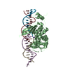
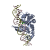
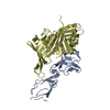
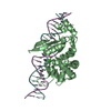
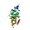
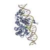
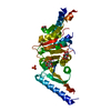
| Title | Structure of E. coli ExoIX in complex with the palindromic 5ov4 DNA oligonucleotide, potassium and calcium | ||||||
 Components Components |
| ||||||
 Keywords Keywords |  HYDROLASE / EXOIX / FLAP ENDONUCLEASE / DNA BINDING HYDROLASE / EXOIX / FLAP ENDONUCLEASE / DNA BINDING | ||||||
| Function / homology |  Function and homology informationDNA replication, Okazaki fragment processing / 5'-flap endonuclease activity / potassium ion binding / Hydrolases; Acting on ester bonds / magnesium ion binding / DNA binding Function and homology informationDNA replication, Okazaki fragment processing / 5'-flap endonuclease activity / potassium ion binding / Hydrolases; Acting on ester bonds / magnesium ion binding / DNA bindingSimilarity search - Function | ||||||
| Biological species |  ESCHERICHIA COLI (E. coli) ESCHERICHIA COLI (E. coli)SYNTHETIC CONSTRUCT (others) | ||||||
| Method | X-RAY DIFFRACTION / SYNCHROTRON / MOLECULAR REPLACEMENT / Resolution: 1.53 Å | ||||||
 Authors Authors | Hemsworth, G.R. / Anstey-Gilbert, C.S. / Flemming, C.S. / Hodskinson, M.R.G. / Zhang, J. / Sedelnikova, S.E. / Stillman, T.J. / Sayers, J.R. / Artymiuk, P.J. | ||||||
 Citation Citation | Journal: Nucleic Acids Res. / Year: 2013 Title: The Structure of E. Coli Exoix - Implications for DNA Binding and Catalysis in Flap Endonucleases Authors: Anstey-Gilbert, C.S. / Hemsworth, G.R. / Flemming, C.S. / Hodskinson, M.R.G. / Zhang, J. / Sedelnikova, S.E. / Stillman, T.J. / Sayers, J.R. / Artymiuk, P.J. | ||||||
| History |
|
- Structure visualization
| Structure viewer | Molecule: MolmilJmol/JSmol |
|---|
- Downloads & links
-Download
| PDBx/mmCIF format | 3zdc.cif.gz | 70.2 KB | Display | PDBx/mmCIF format |
|---|---|---|---|---|
| PDB format | pdb3zdc.ent.gz | 53.6 KB | Display | PDB format |
| PDBx/mmJSON format | 3zdc.json.gz | Tree view | PDBx/mmJSON format | |
| Others |  Other downloads Other downloads |
-Validation report
| Arichive directory | https://data.pdbj.org/pub/pdb/validation_reports/zd/3zdcftp://data.pdbj.org/pub/pdb/validation_reports/zd/3zdc | HTTPS FTP |
|---|
-Related structure data
| Related structure data |  3zd8C  3zd9C  3zdaC  3zdbC  3zddC  3zdeC C: citing same article ( |
|---|---|
| Similar structure data |

-Links
 PDBj
PDBj


- Assembly
| Deposited unit | 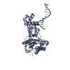
| ||||||||
|---|---|---|---|---|---|---|---|---|---|
| 1 |
| ||||||||
| Unit cell |
|
-Components
-Protein / DNA chain , 2 types, 2 molecules AX
| #1: Protein | Mass: 28203.158 Da / Num. of mol.: 1 Source method: isolated from a genetically manipulated source Source: (gene. exp.) ESCHERICHIA COLI (E. coli) / Strain: XL1 BLUE / Plasmid: PJONEX / Production host: ESCHERICHIA COLI (E. coli) / Strain (production host): BL21(DE3)References: UniProt: Q8X6R9, Hydrolases; Acting on ester bonds |
|---|---|
| #2: DNA chain | Mass: 3680.432 Da / Num. of mol.: 1 / Source method: obtained synthetically / Source: (synth.) SYNTHETIC CONSTRUCT (others) |
-Non-polymers , 4 types, 164 molecules 


| #3: Chemical | ChemComp-K /  Mass: 39.098 Da / Num. of mol.: 1 / Source method: obtained synthetically / Formula: K Mass: 39.098 Da / Num. of mol.: 1 / Source method: obtained synthetically / Formula: K | ||||
|---|---|---|---|---|---|
| #4: Chemical |  Mass: 40.078 Da / Num. of mol.: 2 / Source method: obtained synthetically / Formula: Ca Mass: 40.078 Da / Num. of mol.: 2 / Source method: obtained synthetically / Formula: Ca#5: Chemical | Acetate Mass: 59.044 Da / Num. of mol.: 2 / Source method: obtained synthetically / Formula: C2H3O2 Mass: 59.044 Da / Num. of mol.: 2 / Source method: obtained synthetically / Formula: C2H3O2#6: Water | ChemComp-HOH / | WaterMass: 18.015 Da / Num. of mol.: 159 / Source method: isolated from a natural source / Formula: H2O |
-Details
| Sequence details | OLIGO DESIGNED BASED ON INITIAL STRUCTURE WITH FRAGMENT OF LARGER DNA |
|---|
-Experimental details
-Experiment
| Experiment | Method: X-RAY DIFFRACTION / Number of used crystals: 1 |
|---|
- Sample preparation
| Crystal | Density Matthews: 2.74 Å3/Da / Density % sol: 60 % / Description: NONE |
|---|---|
| Crystal grow | pH: 6.5 / Details: 0.2 M CALCIUM ACETATE, 15% (W/V) PEG-3350, pH 6.5 |
-Data collection
| Diffraction | Mean temperature: 100 K |
|---|---|
| Diffraction source | Source: SYNCHROTRON / Site: Diamond  / Beamline: I03 / Wavelength: 0.98 / Beamline: I03 / Wavelength: 0.98 |
| Detector | Type: ADSC QUANTUM 315 / Detector: CCD / Date: Mar 4, 2009 / Details: MIRRORS |
| Radiation | Monochromator: DOUBLE CRYSTAL / Protocol: SINGLE WAVELENGTH / Monochromatic (M) / Laue (L): M / Scattering type: x-ray |
| Radiation wavelength | Wavelength: 0.98 Å / Relative weight: 1 |
| Reflection | Resolution: 1.53→40 Å / Num. obs: 53386 / % possible obs: 97.9 % / Observed criterion σ(I): 6 / Redundancy: 4.2 % / Rmerge(I) obs: 0.06 / Net I/σ(I): 13.7 |
| Reflection shell | Resolution: 1.53→1.61 Å / Redundancy: 3.4 % / Rmerge(I) obs: 0.42 / Mean I/σ(I) obs: 3.2 / % possible all: 97.9 |
- Processing
| Software |
| ||||||||||||||||||||||||||||||||||||||||||||||||||||||||||||||||||||||||||||||||||||||||||||||||||||||||||||||||||||||||||||||||||||||||||||||||||||||||||||||||||||||||||||||||||||||
|---|---|---|---|---|---|---|---|---|---|---|---|---|---|---|---|---|---|---|---|---|---|---|---|---|---|---|---|---|---|---|---|---|---|---|---|---|---|---|---|---|---|---|---|---|---|---|---|---|---|---|---|---|---|---|---|---|---|---|---|---|---|---|---|---|---|---|---|---|---|---|---|---|---|---|---|---|---|---|---|---|---|---|---|---|---|---|---|---|---|---|---|---|---|---|---|---|---|---|---|---|---|---|---|---|---|---|---|---|---|---|---|---|---|---|---|---|---|---|---|---|---|---|---|---|---|---|---|---|---|---|---|---|---|---|---|---|---|---|---|---|---|---|---|---|---|---|---|---|---|---|---|---|---|---|---|---|---|---|---|---|---|---|---|---|---|---|---|---|---|---|---|---|---|---|---|---|---|---|---|---|---|---|---|
| Refinement | Method to determine structure: MOLECULAR REPLACEMENT Starting model: EXOIX FLAP1 FRAGMENT COMPLEX STRUCTURE Resolution: 1.53→34.53 Å / Cor.coef. Fo:Fc: 0.957 / Cor.coef. Fo:Fc free: 0.946 / SU B: 1.321 / SU ML: 0.049 / Cross valid method: THROUGHOUT / ESU R: 0.076 / ESU R Free: 0.076 / Stereochemistry target values: MAXIMUM LIKELIHOOD Details: HYDROGENS HAVE BEEN ADDED IN THE RIDING POSITIONS. U VALUES REFINED INDIVIDUALLY
| ||||||||||||||||||||||||||||||||||||||||||||||||||||||||||||||||||||||||||||||||||||||||||||||||||||||||||||||||||||||||||||||||||||||||||||||||||||||||||||||||||||||||||||||||||||||
| Solvent computation | Ion probe radii: 0.8 Å / Shrinkage radii: 0.8 Å / VDW probe radii: 1.4 Å / Solvent model: MASK | ||||||||||||||||||||||||||||||||||||||||||||||||||||||||||||||||||||||||||||||||||||||||||||||||||||||||||||||||||||||||||||||||||||||||||||||||||||||||||||||||||||||||||||||||||||||
| Displacement parameters | Biso mean: 24.24 Å2
| ||||||||||||||||||||||||||||||||||||||||||||||||||||||||||||||||||||||||||||||||||||||||||||||||||||||||||||||||||||||||||||||||||||||||||||||||||||||||||||||||||||||||||||||||||||||
| Refinement step | Cycle: LAST / Resolution: 1.53→34.53 Å
| ||||||||||||||||||||||||||||||||||||||||||||||||||||||||||||||||||||||||||||||||||||||||||||||||||||||||||||||||||||||||||||||||||||||||||||||||||||||||||||||||||||||||||||||||||||||
| Refine LS restraints |
|