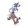
 Movie
Movie Controller


+ Open data

- Basic information
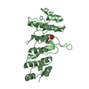
| Entry | Database: PDB / ID: 3wtf | ||||||
|---|---|---|---|---|---|---|---|
| Title | Structure of PAXX | ||||||
 Components Components | Uncharacterized protein C9orf142 | ||||||
 Keywords Keywords |  IMMUNE SYSTEM / DNA repair / Scaffold IMMUNE SYSTEM / DNA repair / Scaffold | ||||||
| Function / homology |  Function and homology informationnonhomologous end joining complex / DNA polymerase binding / double-strand break repair via nonhomologous end joining / site of double-strand break / molecular adaptor activity / DNA damage response / protein homodimerization activity / nucleoplasm / identical protein binding / nucleus / cytoplasm Function and homology informationnonhomologous end joining complex / DNA polymerase binding / double-strand break repair via nonhomologous end joining / site of double-strand break / molecular adaptor activity / DNA damage response / protein homodimerization activity / nucleoplasm / identical protein binding / nucleus / cytoplasmSimilarity search - Function | ||||||
| Biological species |  Homo sapiens (human) Homo sapiens (human) | ||||||
| Method | X-RAY DIFFRACTION / SYNCHROTRON / MOLECULAR REPLACEMENT / Resolution: 3.451 Å | ||||||
 Authors Authors | Ochi, T. / Blundell, T.L. | ||||||
 Citation Citation | Journal: Science / Year: 2015 Title: DNA repair. PAXX, a paralog of XRCC4 and XLF, interacts with Ku to promote DNA double-strand break repair. Authors: Ochi, T. / Blackford, A.N. / Coates, J. / Jhujh, S. / Mehmood, S. / Tamura, N. / Travers, J. / Wu, Q. / Draviam, V.M. / Robinson, C.V. / Blundell, T.L. / Jackson, S.P. | ||||||
| History |
|
- Structure visualization
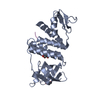
| Structure viewer | Molecule: MolmilJmol/JSmol |
|---|
- Downloads & links
-Download
| PDBx/mmCIF format | 3wtf.cif.gz | 108 KB | Display | PDBx/mmCIF format |
|---|---|---|---|---|
| PDB format | pdb3wtf.ent.gz | 83.6 KB | Display | PDB format |
| PDBx/mmJSON format | 3wtf.json.gz | Tree view | PDBx/mmJSON format | |
| Others |  Other downloads Other downloads |
-Validation report
| Arichive directory | https://data.pdbj.org/pub/pdb/validation_reports/wt/3wtfftp://data.pdbj.org/pub/pdb/validation_reports/wt/3wtf | HTTPS FTP |
|---|
-Related structure data
| Related structure data |  3wtdSC S: Starting model for refinement C: citing same article ( |
|---|---|
| Similar structure data |


-Links
 PDBj
PDBj- Assembly
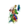
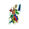
| Deposited unit | 
| ||||||||
|---|---|---|---|---|---|---|---|---|---|
| 1 |
| ||||||||
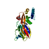
| Unit cell |
|
-Components
| #1: Protein | Mass: 21807.629 Da / Num. of mol.: 2 Source method: isolated from a genetically manipulated source Source: (gene. exp.) Homo sapiens (human) / Gene: C9orf142 / Plasmid: pHAT4 / Production host:  Escherichia coli (E. coli) / Strain (production host): BL21(DE3) / References: UniProt: Q9BUH6 Escherichia coli (E. coli) / Strain (production host): BL21(DE3) / References: UniProt: Q9BUH6#2: Water | ChemComp-HOH / | Water Mass: 18.015 Da / Num. of mol.: 3 / Source method: isolated from a natural source / Formula: H2O Mass: 18.015 Da / Num. of mol.: 3 / Source method: isolated from a natural source / Formula: H2O |
|---|
-Experimental details
-Experiment
| Experiment | Method: X-RAY DIFFRACTION / Number of used crystals: 1 |
|---|
- Sample preparation
| Crystal | Density Matthews: 2.01 Å3/Da / Density % sol: 38.81 % |
|---|---|
| Crystal grow | Temperature: 291 K / Method: vapor diffusion, hanging drop / pH: 6 Details: 1M sodium malonate, 1% (v/v) 1,4-Dioxane, pH 6.0, VAPOR DIFFUSION, HANGING DROP, temperature 291K |
-Data collection
| Diffraction | Mean temperature: 100 K |
|---|---|
| Diffraction source | Source: SYNCHROTRON / Site: Diamond  / Beamline: I03 / Wavelength: 0.97625 Å / Beamline: I03 / Wavelength: 0.97625 Å |
| Detector | Type: DECTRIS PILATUS 6M-F / Detector: PIXEL / Date: May 18, 2013 |
| Radiation | Monochromator: Si 111 / Protocol: SINGLE WAVELENGTH / Monochromatic (M) / Laue (L): M / Scattering type: x-ray |
| Radiation wavelength | Wavelength: 0.97625 Å / Relative weight: 1 |
| Reflection | Resolution: 3.45→24.47 Å / Num. obs: 4986 / % possible obs: 97.3 % / Redundancy: 6.1 % / Rsym value: 0.088 / Net I/σ(I): 9.1 |
| Reflection shell | Resolution: 3.45→3.78 Å / Redundancy: 5.9 % / Mean I/σ(I) obs: 1.7 / Num. unique all: 1150 / Rsym value: 0.599 / % possible all: 98.2 |
- Processing
| Software |
| |||||||||||||||||||||||||||||||||||||||||||||||||||||||||||||||||||||||||||
|---|---|---|---|---|---|---|---|---|---|---|---|---|---|---|---|---|---|---|---|---|---|---|---|---|---|---|---|---|---|---|---|---|---|---|---|---|---|---|---|---|---|---|---|---|---|---|---|---|---|---|---|---|---|---|---|---|---|---|---|---|---|---|---|---|---|---|---|---|---|---|---|---|---|---|---|---|
| Refinement | Method to determine structure: MOLECULAR REPLACEMENT Starting model: 3WTD Resolution: 3.451→24.47 Å / SU ML: 0.52 / σ(F): 1.34 / Phase error: 35.76 / Stereochemistry target values: ML
| |||||||||||||||||||||||||||||||||||||||||||||||||||||||||||||||||||||||||||
| Solvent computation | Shrinkage radii: 0.9 Å / VDW probe radii: 1.11 Å / Solvent model: FLAT BULK SOLVENT MODEL | |||||||||||||||||||||||||||||||||||||||||||||||||||||||||||||||||||||||||||
| Refinement step | Cycle: LAST / Resolution: 3.451→24.47 Å
| |||||||||||||||||||||||||||||||||||||||||||||||||||||||||||||||||||||||||||
| Refine LS restraints |
| |||||||||||||||||||||||||||||||||||||||||||||||||||||||||||||||||||||||||||
| LS refinement shell | Refine-ID: X-RAY DIFFRACTION / Total num. of bins used: 4
| |||||||||||||||||||||||||||||||||||||||||||||||||||||||||||||||||||||||||||
| Refinement TLS params. | Method: refined / Refine-ID: X-RAY DIFFRACTION
| |||||||||||||||||||||||||||||||||||||||||||||||||||||||||||||||||||||||||||
| Refinement TLS group |
|
