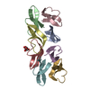
 Movie
Movie Controller


+ Open data

- Basic information
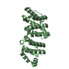
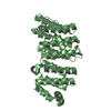
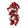
| Entry | Database: PDB / ID: 3woy | ||||||
|---|---|---|---|---|---|---|---|
| Title | Crystal structure of CLASP2 TOG domain (TOG2) | ||||||
 Components Components | CLIP-associating protein 2 | ||||||
 Keywords Keywords |  STRUCTURAL PROTEIN / HEAT Repeat / Microtubule binding / Tubulin / unknown / microtubule STRUCTURAL PROTEIN / HEAT Repeat / Microtubule binding / Tubulin / unknown / microtubule | ||||||
| Function / homology |  Function and homology information Function and homology informationcortical microtubule plus-end / positive regulation of basement membrane assembly involved in embryonic body morphogenesis / negative regulation of wound healing, spreading of epidermal cells / regulation of gastrulation / vesicle targeting / microtubule anchoring / basal cortex / positive regulation of extracellular matrix disassembly / microtubule organizing center organization / kinetochore microtubule ...cortical microtubule plus-end / positive regulation of basement membrane assembly involved in embryonic body morphogenesis / negative regulation of wound healing, spreading of epidermal cells / regulation of gastrulation / vesicle targeting / microtubule anchoring / basal cortex / positive regulation of extracellular matrix disassembly / microtubule organizing center organization / kinetochore microtubule / establishment of mitotic spindle localization / Role of ABL in ROBO-SLIT signaling / dystroglycan binding / regulation of epithelial to mesenchymal transition / negative regulation of focal adhesion assembly / microtubule nucleation / negative regulation of microtubule depolymerization / microtubule plus-end binding / exit from mitosis / regulation of microtubule-based process / regulation of axon extension / microtubule organizing center / negative regulation of stress fiber assembly / establishment or maintenance of cell polarity / positive regulation of epithelial cell migration / regulation of microtubule polymerization / Golgi organization / positive regulation of exocytosis / cell leading edge / platelet-derived growth factor receptor-beta signaling pathway / mitotic spindle assembly / regulation of microtubule polymerization or depolymerization / cytoplasmic microtubule / Amplification of signal from unattached kinetochores via a MAD2 inhibitory signal / axonal growth cone / Mitotic Prometaphase / EML4 and NUDC in mitotic spindle formation / Resolution of Sister Chromatid Cohesion / protein tyrosine kinase binding / mitotic spindle organization / RHO GTPases Activate Formins / spindle microtubule / protein localization to plasma membrane / regulation of actin cytoskeleton organization / trans-Golgi network / microtubule cytoskeleton organization / kinetochore / ruffle membrane / Separation of Sister Chromatids / actin filament binding / cell cortex / microtubule binding / microtubule / cell division / centrosome / Golgi apparatus / membrane / plasma membrane / cytosol / cytoplasmSimilarity search - Function | ||||||
| Biological species |  Homo sapiens (human) Homo sapiens (human) | ||||||
| Method | X-RAY DIFFRACTION / SYNCHROTRON / MAD / Resolution: 2.1 Å | ||||||
 Authors Authors | Hayashi, I. / Maki, T. | ||||||
 Citation Citation | Journal: J. Mol. Biol. / Year: 2015 Title: CLASP2 Has Two Distinct TOG Domains That Contribute Differently to Microtubule Dynamics Authors: Maki, T. / Grimaldi, A.D. / Fuchigami, S. / Kaverina, I. / Hayashi, I. | ||||||
| History |
|
- Structure visualization
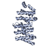
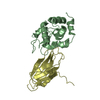
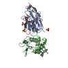
| Structure viewer | Molecule: MolmilJmol/JSmol |
|---|
- Downloads & links
-Download
| PDBx/mmCIF format | 3woy.cif.gz | 65.1 KB | Display | PDBx/mmCIF format |
|---|---|---|---|---|
| PDB format | pdb3woy.ent.gz | 47.4 KB | Display | PDB format |
| PDBx/mmJSON format | 3woy.json.gz | Tree view | PDBx/mmJSON format | |
| Others |  Other downloads Other downloads |
-Validation report
| Arichive directory | https://data.pdbj.org/pub/pdb/validation_reports/wo/3woyftp://data.pdbj.org/pub/pdb/validation_reports/wo/3woy | HTTPS FTP |
|---|
-Related structure data


-Links
 PDBj
PDBj


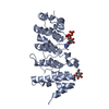
- Assembly
| Deposited unit | 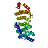
| ||||||||
|---|---|---|---|---|---|---|---|---|---|
| 1 |
| ||||||||
| Unit cell |
|
-Components
| #1: Protein | Mass: 28182.201 Da / Num. of mol.: 1 / Fragment: UNP residues 60-310 Source method: isolated from a genetically manipulated source Source: (gene. exp.) Homo sapiens (human) / Gene: CLASP2, KIAA0627 / Production host:  Escherichia coli (E. coli) / References: UniProt: O75122 Escherichia coli (E. coli) / References: UniProt: O75122 |
|---|---|
| #2: Water | ChemComp-HOH / Water Mass: 18.015 Da / Num. of mol.: 212 / Source method: isolated from a natural source / Formula: H2O Mass: 18.015 Da / Num. of mol.: 212 / Source method: isolated from a natural source / Formula: H2O |
-Experimental details
-Experiment
| Experiment | Method: X-RAY DIFFRACTION / Number of used crystals: 1 |
|---|
- Sample preparation
| Crystal | Density Matthews: 2.78 Å3/Da / Density % sol: 55.8 % |
|---|---|
| Crystal grow | Temperature: 293 K / Method: vapor diffusion, hanging drop / pH: 8.5 Details: 20mM Tris, 3%(w/v) PEG10K , pH 8.5, VAPOR DIFFUSION, HANGING DROP, temperature 293K |
-Data collection
| Diffraction |
| |||||||||||||||||||||||||||||||||
|---|---|---|---|---|---|---|---|---|---|---|---|---|---|---|---|---|---|---|---|---|---|---|---|---|---|---|---|---|---|---|---|---|---|---|
| Diffraction source |
| |||||||||||||||||||||||||||||||||
| Detector |
| |||||||||||||||||||||||||||||||||
| Radiation |
| |||||||||||||||||||||||||||||||||
| Radiation wavelength |
| |||||||||||||||||||||||||||||||||
| Reflection | Resolution: 2→50 Å / Num. obs: 44169 / % possible obs: 99.9 % / Observed criterion σ(F): 62 / Observed criterion σ(I): 2.9 / Biso Wilson estimate: 25.4 Å2 | |||||||||||||||||||||||||||||||||
| Reflection shell |
|

- Processing
| Software |
| ||||||||||||||||||||||||||||||||||||||||||||||||||||||||||||
|---|---|---|---|---|---|---|---|---|---|---|---|---|---|---|---|---|---|---|---|---|---|---|---|---|---|---|---|---|---|---|---|---|---|---|---|---|---|---|---|---|---|---|---|---|---|---|---|---|---|---|---|---|---|---|---|---|---|---|---|---|---|
| Refinement | Method to determine structure: MAD / Resolution: 2.1→42.73 Å / Rfactor Rfree error: 0.006 / Data cutoff high absF: 172351.58 / Data cutoff low absF: 0 / Isotropic thermal model: RESTRAINED / Cross valid method: THROUGHOUT / σ(F): 0 / Details: BULK SOLVENT MODEL USED
| ||||||||||||||||||||||||||||||||||||||||||||||||||||||||||||
| Solvent computation | Solvent model: FLAT MODEL / Bsol: 59.0081 Å2 / ksol: 0.36 e/Å3 | ||||||||||||||||||||||||||||||||||||||||||||||||||||||||||||
| Displacement parameters | Biso mean: 42.7 Å2
| ||||||||||||||||||||||||||||||||||||||||||||||||||||||||||||
| Refine analyze |
| ||||||||||||||||||||||||||||||||||||||||||||||||||||||||||||
| Refinement step | Cycle: LAST / Resolution: 2.1→42.73 Å
| ||||||||||||||||||||||||||||||||||||||||||||||||||||||||||||
| Refine LS restraints |
| ||||||||||||||||||||||||||||||||||||||||||||||||||||||||||||
| LS refinement shell | Resolution: 2.1→2.23 Å / Rfactor Rfree error: 0.017 / Total num. of bins used: 6
| ||||||||||||||||||||||||||||||||||||||||||||||||||||||||||||
| Xplor file |
|
