 Movie
Movie Controller

[English] 日本語
 Yorodumi
- PDB-3wkm: The periplasmic PDZ tandem fragment of the RseP homologue from Aq... -
+ Open data

- Basic information
| Entry | Database: PDB / ID: 3wkm | ||||||
|---|---|---|---|---|---|---|---|
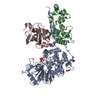
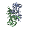
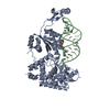
| Title | The periplasmic PDZ tandem fragment of the RseP homologue from Aquifex aeolicus in complex with the Fab fragment | ||||||
 Components Components |
| ||||||
 Keywords Keywords |  HYDROLASE / PDZ DOMAIN HYDROLASE / PDZ DOMAIN | ||||||
| Function / homology |  Function and homology informationHydrolases; Acting on peptide bonds (peptidases); Metalloendopeptidases / metalloendopeptidase activity / proteolysis / metal ion binding / plasma membrane Function and homology informationHydrolases; Acting on peptide bonds (peptidases); Metalloendopeptidases / metalloendopeptidase activity / proteolysis / metal ion binding / plasma membraneSimilarity search - Function | ||||||
| Biological species |   Aquifex aeolicus (bacteria) Aquifex aeolicus (bacteria) MUS MUSCULUS (house mouse) MUS MUSCULUS (house mouse) | ||||||
| Method | X-RAY DIFFRACTION / SYNCHROTRON / MOLECULAR REPLACEMENT / Resolution: 2.2 Å | ||||||
 Authors Authors | Nogi, T. / Tabata, S. / Tamura-kawakami, K. / Takagi, J. | ||||||
 Citation Citation | Journal: Structure / Year: 2013 Title: A Structure-Based Model of Substrate Discrimination by a Noncanonical PDZ Tandem in the Intramembrane-Cleaving Protease RseP Authors: Hizukuri, Y. / Oda, T. / Tabata, S. / Tamura-kawakami, K. / Oi, R. / Sato, M. / Takagi, J. / Akiyama, Y. / Nogi, T. | ||||||
| History |
|
- Structure visualization
| Structure viewer | Molecule: MolmilJmol/JSmol |
|---|
- Downloads & links
-Download
| PDBx/mmCIF format | 3wkm.cif.gz | 250.1 KB | Display | PDBx/mmCIF format |
|---|---|---|---|---|
| PDB format | pdb3wkm.ent.gz | 199.6 KB | Display | PDB format |
| PDBx/mmJSON format | 3wkm.json.gz | Tree view | PDBx/mmJSON format | |
| Others |  Other downloads Other downloads |
-Validation report
| Arichive directory | https://data.pdbj.org/pub/pdb/validation_reports/wk/3wkmftp://data.pdbj.org/pub/pdb/validation_reports/wk/3wkm | HTTPS FTP |
|---|
-Related structure data
| Related structure data |  3wklC  1nsnS C: citing same article ( S: Starting model for refinement |
|---|---|
| Similar structure data |


-Links
 PDBj
PDBj


- Assembly
| Deposited unit | 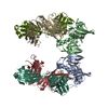
| ||||||||
|---|---|---|---|---|---|---|---|---|---|
| 1 | 
| ||||||||
| 2 | 
| ||||||||
| Unit cell |
|
-Components
| #1: Protein | Mass: 19997.455 Da / Num. of mol.: 2 / Fragment: PDZ TANDEM FRAGMENT, UNP RESIDUES 115-292 Source method: isolated from a genetically manipulated source Source: (gene. exp.) Aquifex aeolicus (bacteria) / Strain: VF5 / Gene: aq_1964 / Production host: ESCHERICHIA COLI (E. coli)References: UniProt: O67776, Hydrolases; Acting on peptide bonds (peptidases); Metalloendopeptidases#2: Antibody | Mass: 24268.041 Da / Num. of mol.: 2 / Source method: isolated from a natural source / Source: (natural) MUS MUSCULUS (house mouse)#3: Antibody | Mass: 24080.465 Da / Num. of mol.: 2 / Source method: isolated from a natural source / Source: (natural) MUS MUSCULUS (house mouse)#4: Water | ChemComp-HOH / | Water Mass: 18.015 Da / Num. of mol.: 387 / Source method: isolated from a natural source / Formula: H2O Mass: 18.015 Da / Num. of mol.: 387 / Source method: isolated from a natural source / Formula: H2OSequence details | DNA SEQUENCEs FOR CHAIN H, I, L AND M WERE NOT AVAILABLE AT THE UNIPROT KNOWLEDGEBASE DATABASE ...DNA SEQUENCEs FOR CHAIN H, I, L AND M WERE NOT AVAILABLE AT THE UNIPROT KNOWLEDGEB | |
|---|
-Experimental details
-Experiment
| Experiment | Method: X-RAY DIFFRACTION / Number of used crystals: 1 |
|---|
- Sample preparation
| Crystal | Density Matthews: 2.84 Å3/Da / Density % sol: 56.63 % |
|---|---|
| Crystal grow | pH: 7 Details: 16%(W/V) POLYETHYLENE GLYCOL 8000, 200MM SODIUM MALONATE, 0.5%(W/) N-OCTYL BETA-D-GLUCOPYRANOSIDE, PH 7.0, VAPOR DIFFUSION, SITTING DROP, TEMPERATURE 293K |
-Data collection
| Diffraction | Mean temperature: 100 K |
|---|---|
| Diffraction source | Source: SYNCHROTRON / Site: Photon Factory  / Beamline: BL-17A / Wavelength: 1 / Beamline: BL-17A / Wavelength: 1 |
| Detector | Type: ADSC QUANTUM 270 / Detector: CCD / Date: Jun 10, 2011 |
| Radiation | Monochromator: SI(111) / Protocol: SINGLE WAVELENGTH / Monochromatic (M) / Laue (L): M / Scattering type: x-ray |
| Radiation wavelength | Wavelength: 1 Å / Relative weight: 1 |
| Reflection | Resolution: 2.2→47.44 Å / Num. obs: 74923 / % possible obs: 98.2 % / Observed criterion σ(I): -3 |
| Reflection shell | Resolution: 2.2→2.32 Å / % possible all: 97.2 |
- Processing
| Software |
| ||||||||||||||||||||||||||||||||||||||||||||||||||||||||||||||||||||||||||||||||||||||||||||||||||||||||||||||||||||||||||||||||||||||||||||||||||||||||||||||||||||||||||||||||||||||
|---|---|---|---|---|---|---|---|---|---|---|---|---|---|---|---|---|---|---|---|---|---|---|---|---|---|---|---|---|---|---|---|---|---|---|---|---|---|---|---|---|---|---|---|---|---|---|---|---|---|---|---|---|---|---|---|---|---|---|---|---|---|---|---|---|---|---|---|---|---|---|---|---|---|---|---|---|---|---|---|---|---|---|---|---|---|---|---|---|---|---|---|---|---|---|---|---|---|---|---|---|---|---|---|---|---|---|---|---|---|---|---|---|---|---|---|---|---|---|---|---|---|---|---|---|---|---|---|---|---|---|---|---|---|---|---|---|---|---|---|---|---|---|---|---|---|---|---|---|---|---|---|---|---|---|---|---|---|---|---|---|---|---|---|---|---|---|---|---|---|---|---|---|---|---|---|---|---|---|---|---|---|---|---|
| Refinement | Method to determine structure: MOLECULAR REPLACEMENT Starting model: 1NSN Resolution: 2.2→47.44 Å / Cor.coef. Fo:Fc: 0.931 / Cor.coef. Fo:Fc free: 0.896 / SU B: 6.989 / SU ML: 0.177 / Cross valid method: THROUGHOUT / ESU R: 0.279 / ESU R Free: 0.227 / Stereochemistry target values: MAXIMUM LIKELIHOOD / Details: HYDROGENS HAVE BEEN USED IF PRESENT IN THE INPUT
| ||||||||||||||||||||||||||||||||||||||||||||||||||||||||||||||||||||||||||||||||||||||||||||||||||||||||||||||||||||||||||||||||||||||||||||||||||||||||||||||||||||||||||||||||||||||
| Solvent computation | Ion probe radii: 0.8 Å / Shrinkage radii: 0.8 Å / VDW probe radii: 1.2 Å / Solvent model: MASK | ||||||||||||||||||||||||||||||||||||||||||||||||||||||||||||||||||||||||||||||||||||||||||||||||||||||||||||||||||||||||||||||||||||||||||||||||||||||||||||||||||||||||||||||||||||||
| Displacement parameters | Biso mean: 39.78 Å2
| ||||||||||||||||||||||||||||||||||||||||||||||||||||||||||||||||||||||||||||||||||||||||||||||||||||||||||||||||||||||||||||||||||||||||||||||||||||||||||||||||||||||||||||||||||||||
| Refinement step | Cycle: LAST / Resolution: 2.2→47.44 Å
| ||||||||||||||||||||||||||||||||||||||||||||||||||||||||||||||||||||||||||||||||||||||||||||||||||||||||||||||||||||||||||||||||||||||||||||||||||||||||||||||||||||||||||||||||||||||
| Refine LS restraints |
|