 Movie
Movie Controller

[English] 日本語
 Yorodumi
- PDB-3wcz: Crystal structure of Bombyx mori aldo-keto reductase (AKR2E4) in ... -
+ Open data

- Basic information
| Entry | Database: PDB / ID: 3wcz | ||||||
|---|---|---|---|---|---|---|---|
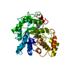
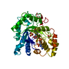
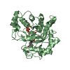
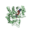
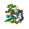
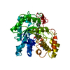
| Title | Crystal structure of Bombyx mori aldo-keto reductase (AKR2E4) in complex with NADP | ||||||
 Components Components | Aldo-keto reductase 2E | ||||||
 Keywords Keywords |  OXIDOREDUCTASE / TIM-BARREL / REDUCTASE / NADP / CYTOSOL OXIDOREDUCTASE / TIM-BARREL / REDUCTASE / NADP / CYTOSOL | ||||||
| Function / homology |  Function and homology informationOxidoreductases; Acting on the CH-OH group of donors; With NAD+ or NADP+ as acceptor / steroid metabolic process / oxidoreductase activity Function and homology informationOxidoreductases; Acting on the CH-OH group of donors; With NAD+ or NADP+ as acceptor / steroid metabolic process / oxidoreductase activitySimilarity search - Function | ||||||
| Biological species |  Bombyx mori (domestic silkworm) Bombyx mori (domestic silkworm) | ||||||
| Method | X-RAY DIFFRACTION / SYNCHROTRON / MOLECULAR REPLACEMENT / Resolution: 1.3 Å | ||||||
 Authors Authors | Yamamoto, K. / Wilson, D.K. | ||||||
 Citation Citation | Journal: Arch.Biochem.Biophys. / Year: 2013 Title: Identification, characterization, and crystal structure of an aldo-keto reductase (AKR2E4) from the silkworm Bombyx mori. Authors: Yamamoto, K. / Wilson, D.K. | ||||||
| History |
|
- Structure visualization
| Structure viewer | Molecule: MolmilJmol/JSmol |
|---|
- Downloads & links
-Download
| PDBx/mmCIF format | 3wcz.cif.gz | 133.8 KB | Display | PDBx/mmCIF format |
|---|---|---|---|---|
| PDB format | pdb3wcz.ent.gz | 104.9 KB | Display | PDB format |
| PDBx/mmJSON format | 3wcz.json.gz | Tree view | PDBx/mmJSON format | |
| Others |  Other downloads Other downloads |
-Validation report
| Arichive directory | https://data.pdbj.org/pub/pdb/validation_reports/wc/3wczftp://data.pdbj.org/pub/pdb/validation_reports/wc/3wcz | HTTPS FTP |
|---|
-Related structure data
| Related structure data |  2hdjS S: Starting model for refinement |
|---|---|
| Similar structure data |

-Links
 PDBj
PDBj
- Assembly
| Deposited unit | 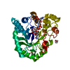
| ||||||||
|---|---|---|---|---|---|---|---|---|---|
| 1 |
| ||||||||
| Unit cell |
|
-Components
| #1: Protein | Mass: 34385.652 Da / Num. of mol.: 1 Source method: isolated from a genetically manipulated source Source: (gene. exp.) Bombyx mori (domestic silkworm) / Gene: akr2e / Production host:  Escherichia coli (E. coli) / References: UniProt: H9JTG9 Escherichia coli (E. coli) / References: UniProt: H9JTG9 | ||||
|---|---|---|---|---|---|
| #2: Chemical | ChemComp-NAP / Nicotinamide adenine dinucleotide phosphate  Mass: 743.405 Da / Num. of mol.: 1 / Source method: obtained synthetically / Formula: C21H28N7O17P3 Mass: 743.405 Da / Num. of mol.: 1 / Source method: obtained synthetically / Formula: C21H28N7O17P3 | ||||
| #3: Chemical | ChemComp-EDO / Ethylene glycol  Mass: 62.068 Da / Num. of mol.: 4 / Source method: obtained synthetically / Formula: C2H6O2 Mass: 62.068 Da / Num. of mol.: 4 / Source method: obtained synthetically / Formula: C2H6O2#4: Chemical | ChemComp-CIT / | Citric acid  Mass: 192.124 Da / Num. of mol.: 1 / Source method: obtained synthetically / Formula: C6H8O7 Mass: 192.124 Da / Num. of mol.: 1 / Source method: obtained synthetically / Formula: C6H8O7#5: Water | ChemComp-HOH / | Water Mass: 18.015 Da / Num. of mol.: 237 / Source method: isolated from a natural source / Formula: H2O Mass: 18.015 Da / Num. of mol.: 237 / Source method: isolated from a natural source / Formula: H2O |
-Experimental details
-Experiment
| Experiment | Method: X-RAY DIFFRACTION / Number of used crystals: 1 |
|---|
- Sample preparation
| Crystal | Density Matthews: 2.1 Å3/Da / Density % sol: 41.5 % |
|---|---|
| Crystal grow | Temperature: 295.15 K / Method: vapor diffusion, hanging drop / pH: 6.5 Details: 1M SODIUM CITRATE, 0.1M CACODYLATE (PH 6.5), VAPOR DIFFUSION, HANGING DROP, TEMPERATURE 295.15K |
-Data collection
| Diffraction | Mean temperature: 100 K |
|---|---|
| Diffraction source | Source: SYNCHROTRON / Site: SSRL  / Beamline: BL9-2 / Wavelength: 0.98 / Beamline: BL9-2 / Wavelength: 0.98 |
| Detector | Type: MARMOSAIC 325 mm CCD / Detector: CCD |
| Radiation | Protocol: SINGLE WAVELENGTH / Monochromatic (M) / Laue (L): M / Scattering type: x-ray |
| Radiation wavelength | Wavelength: 0.98 Å / Relative weight: 1 |
| Reflection | Resolution: 1.3→34.503 Å / Num. obs: 69041 / % possible obs: 98.5 % / Observed criterion σ(I): 3 |
| Reflection shell | Highest resolution: 1.3 Å / % possible all: 100 |
- Processing
| Software |
| ||||||||||||||||||||||||||||||||||||||||||||||||||||||||||||||||||||||||||||||||||||||||||
|---|---|---|---|---|---|---|---|---|---|---|---|---|---|---|---|---|---|---|---|---|---|---|---|---|---|---|---|---|---|---|---|---|---|---|---|---|---|---|---|---|---|---|---|---|---|---|---|---|---|---|---|---|---|---|---|---|---|---|---|---|---|---|---|---|---|---|---|---|---|---|---|---|---|---|---|---|---|---|---|---|---|---|---|---|---|---|---|---|---|---|---|
| Refinement | Method to determine structure: MOLECULAR REPLACEMENT Starting model: 2HDJ Resolution: 1.3→34.5 Å / SU ML: 0.23 / σ(F): 1.34 / Phase error: 16.65 / Stereochemistry target values: MLHL
| ||||||||||||||||||||||||||||||||||||||||||||||||||||||||||||||||||||||||||||||||||||||||||
| Solvent computation | Shrinkage radii: 0.17 Å / VDW probe radii: 0.4 Å / Solvent model: FLAT BULK SOLVENT MODEL / Bsol: 45 Å2 / ksol: 0.5 e/Å3 | ||||||||||||||||||||||||||||||||||||||||||||||||||||||||||||||||||||||||||||||||||||||||||
| Displacement parameters |
| ||||||||||||||||||||||||||||||||||||||||||||||||||||||||||||||||||||||||||||||||||||||||||
| Refinement step | Cycle: LAST / Resolution: 1.3→34.5 Å
| ||||||||||||||||||||||||||||||||||||||||||||||||||||||||||||||||||||||||||||||||||||||||||
| Refine LS restraints |
| ||||||||||||||||||||||||||||||||||||||||||||||||||||||||||||||||||||||||||||||||||||||||||
| LS refinement shell | Refine-ID: X-RAY DIFFRACTION / Total num. of bins used: 14
|
